Galeazzi fractures
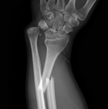
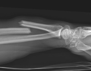
Definition
Fracture of the radial shaft with disruption to the distal radio-ulna joint (DRUJ)


Fracture of the radial shaft with disruption to the distal radio-ulna joint (DRUJ)
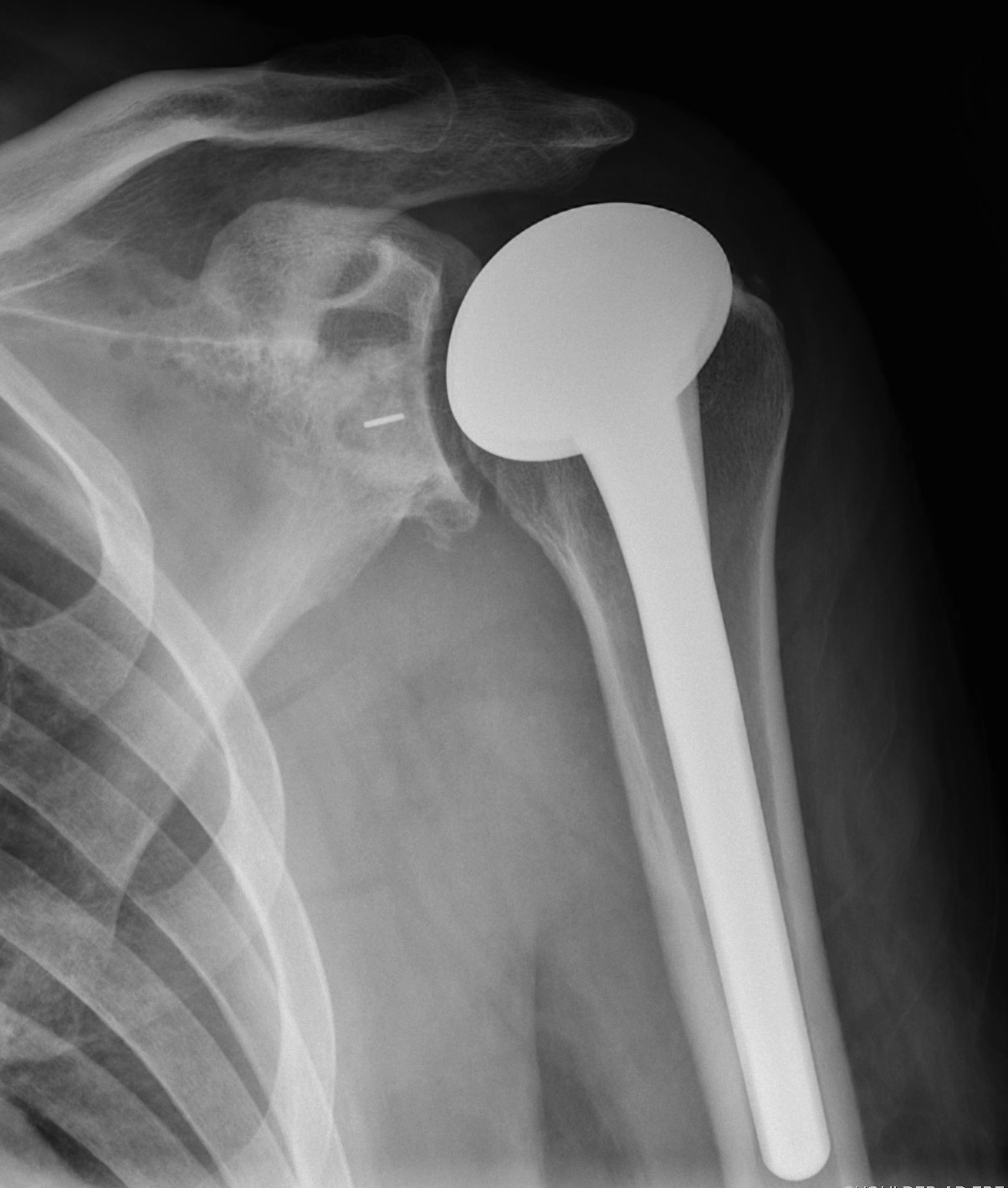
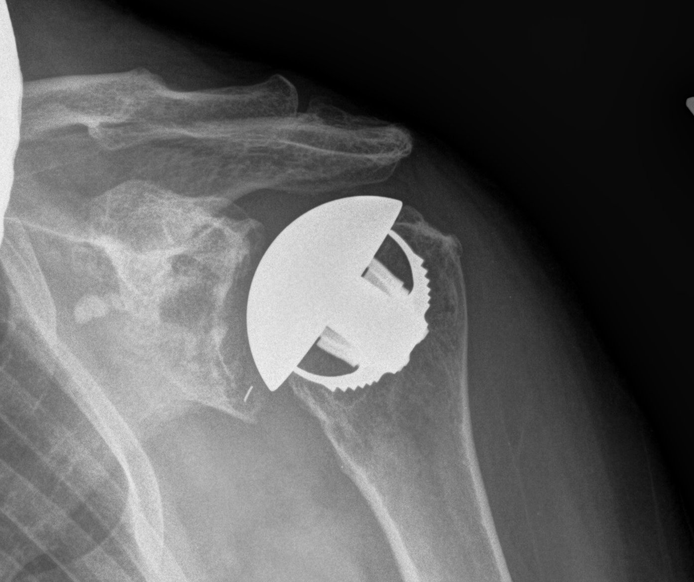
Osteoarthritis
Rheumatoid arthritis
Avascular necrosis
1. Significant functional impairment
2. PIPJ contracture
- originally thought to intervene early
- Macfarlane showed residual FFD always about 30o
- may need to release check rein ligaments / accessory collateral ligaments
3. MCPJ contracture >30o
4. Trigger fingers
- must do limited fasciectomy
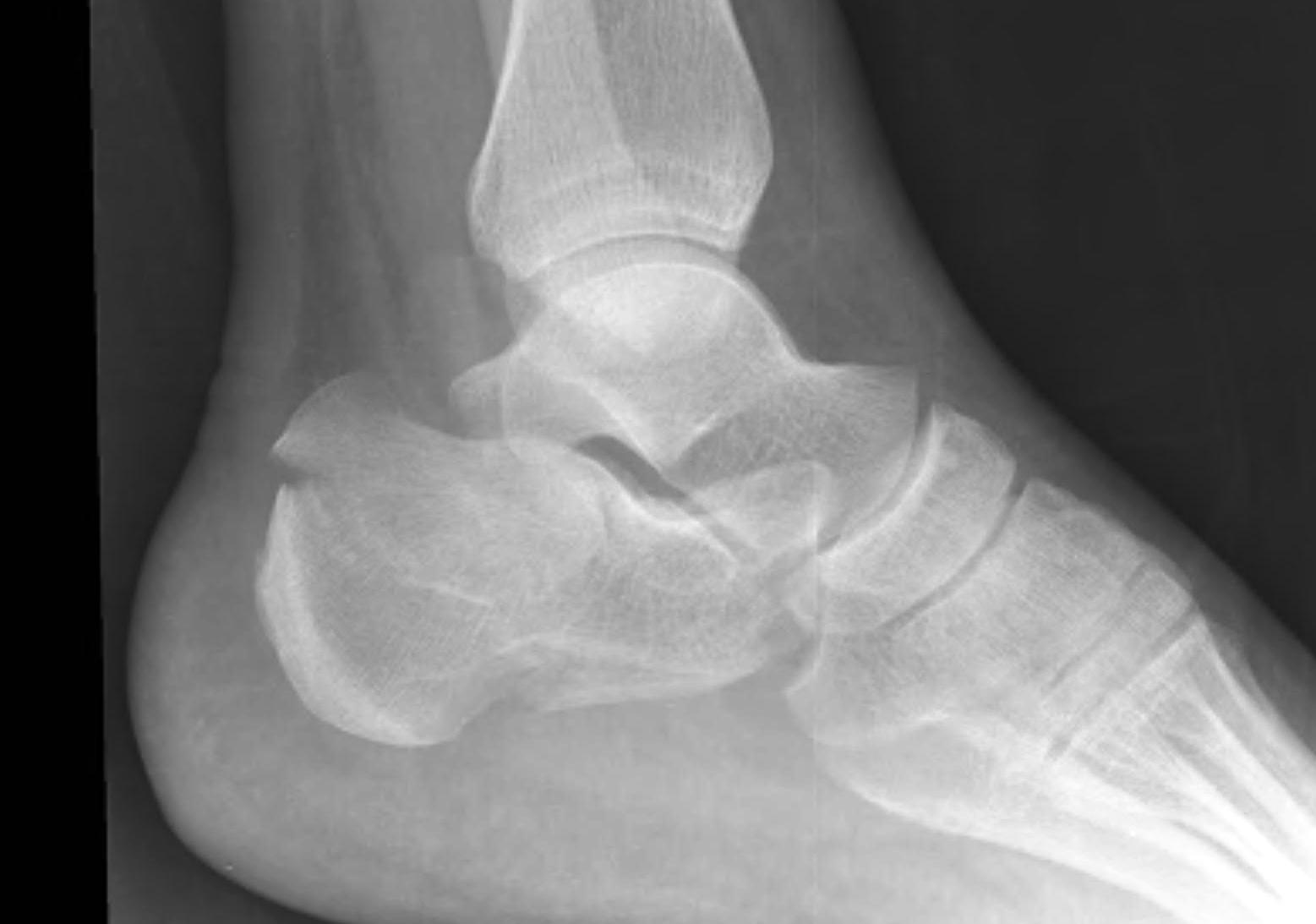
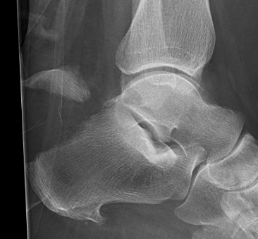
Osseous canal between talus and calcaneum
- interosseous talo-calcaneal ligament
- cervical ligament
- joint capsule
- nerve endings / arterial anastomoses
Flat foot / overpronation
Inversion / sprain
Proximal tibia
- gastrocnemius local muscle flap
- gracilis free muscle flap if gastroc damaged
Middle tibia
- soleus local muscle flap
- gracilis free muscle flap
Distal tibia
- posterior tibial fasciocutaneous local flap
- gracilis free muscle flap
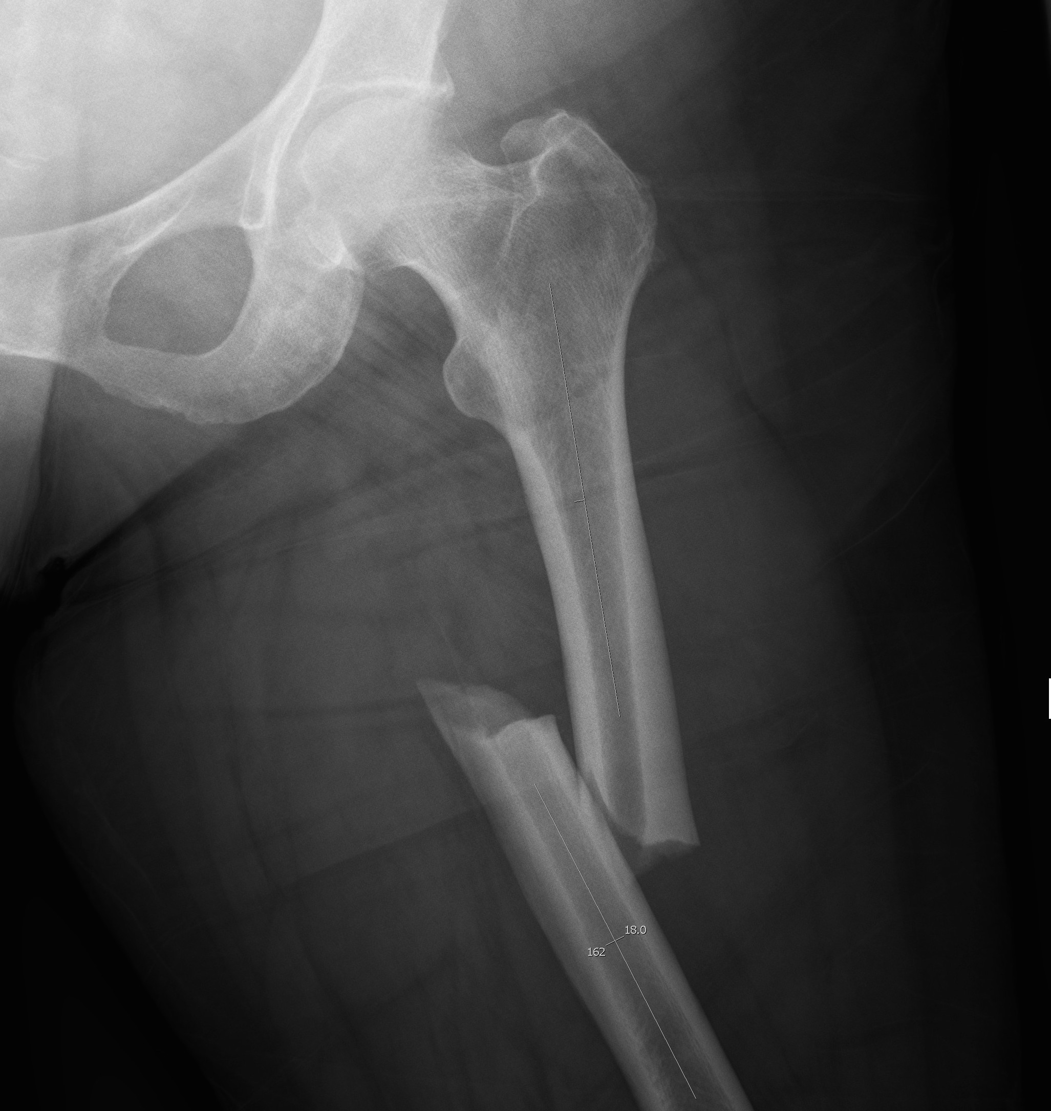
Usually young patients
- 15 - 40
15% compound
High velocity injury
- MBA
- MVA
- pedestrian v car
- fall from height
EMST principles
- need for transfusion not uncommon
1. Open antero-lateral approach
Large / Massive Cuff Tear
2. Deltopectoral approach
Large Subscapularis tear
3. Arthroscopic Assisted Mini-open
Indication
- Small / Moderate Cuff Tear < 3cm
- no retraction
Technique
- arthroscopic SAD
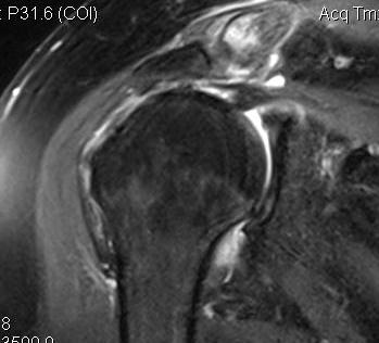
Massive tear
1. > 5cm
- retracted to humerus / glenoid margin
2. At least 2 complete tendons
- lose SS / IS or SS / SC