xray

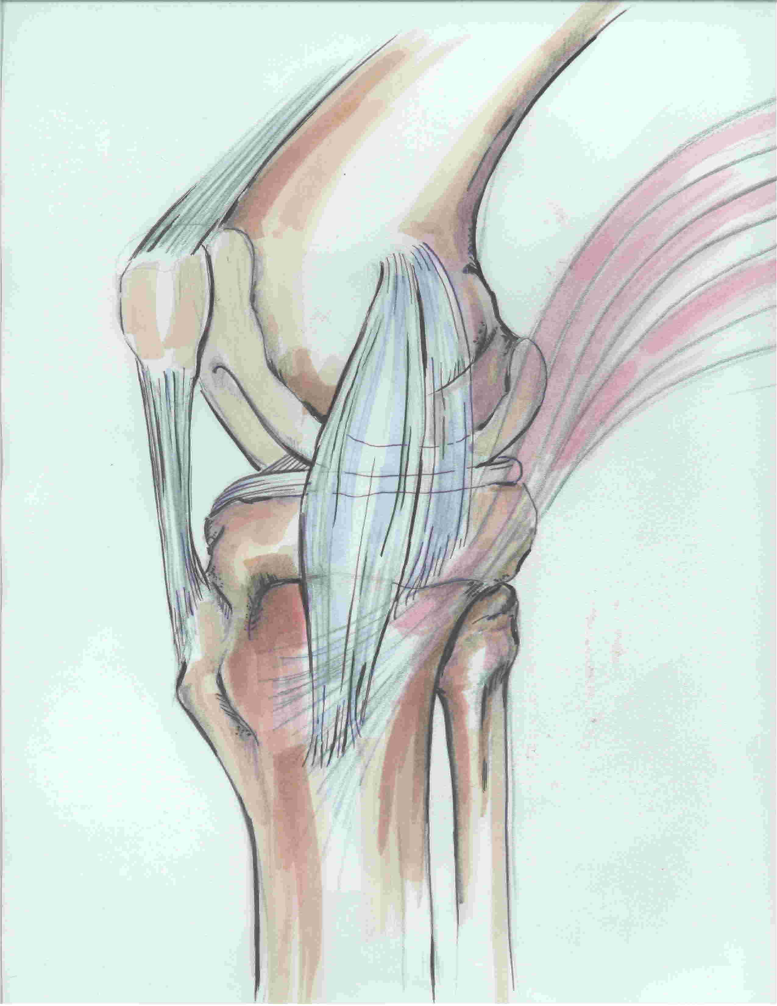
Patella Tendon Rupture

Epidemiology


Usually occurs in young people

- often previous history of tendonitis ± steroid injections
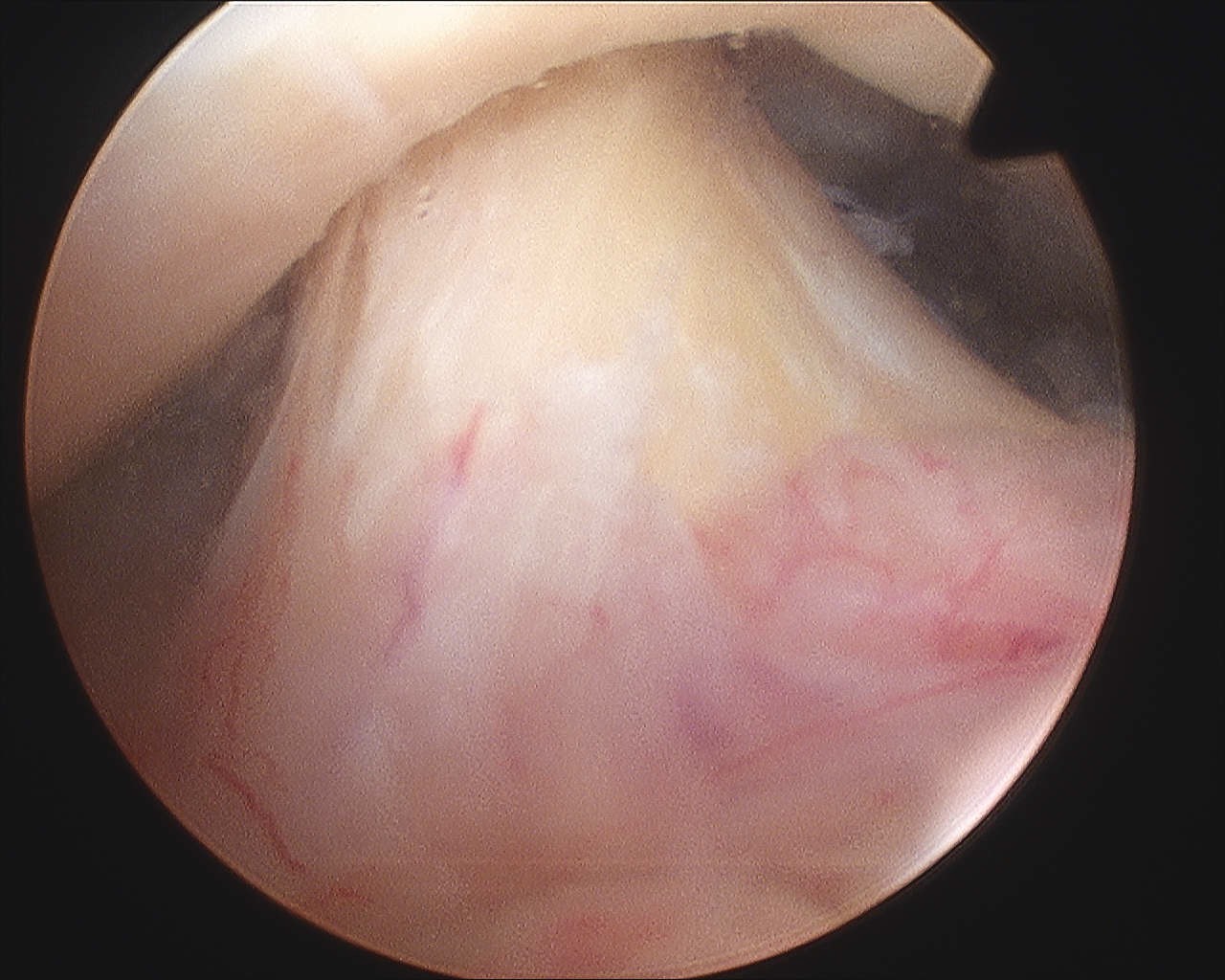
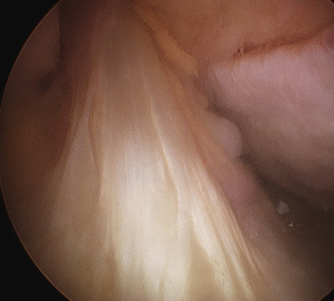
 

Location


Usually at level of inferior pole of patella

- less common at tibial tubercle

- mid-substance ruptures rare


Clinical


Severe pain

Palpable defect

Extensor deficit / unable to SLR
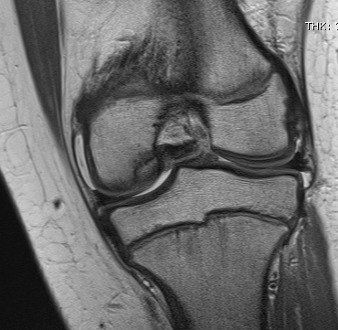
 

Xray


Patella alta / high riding patella


Background

DefinitionKnee OCD MRI Cartilage Intact


Osteochondritis Dissecans

- separation of avascular fragment of bone & cartilage


Epidemiology


M : F = 2:1


Mean age 18 years

- can present as young as 9


4:1000


Background


ACL Normal ArthroscopyACL Normal Arthroscopy


Anatomy


Developmental Anatomy


Knee joint first appears as a mesenchymal cleft at 8 weeks gestation

- ACL and PCL separate entities by week 10

MCL and Posteromedial Corner

AnatomyMCL anatomy


1. Seebacher's 3 layers of the medial knee


Layer 1

- sartorius and sartorius fascia


Layer 2

- superficial MCL

- posterior oblique ligament

- semimembranosus


Layer 3

- deep MCL (meniscofemoral and meniscotibial ligament)

- posteromedial capsule 


2. MCL


Superficial MCL

AVN


SONK              vs                          Atraumatic AVN


>55                                              Often mid 30's

Templating

AimTHR Templating


Reproduce the normal anatomical centre of rotation

Restore femoral offset 

Maintain equal leg lengths 


Usually template off normal hip


Template


1. LLD

2. Offset

3. Femoral component

4. Acetabular component

5. Osteotomy / femoral seating


Background

IssuesTHR Uncemented


Templating

Approach

Fixation

Bearing Surface

Head Size

Offset


Indications


Disabling hip pain

Severe functional impairment

Failed non operative management


Not Indicated 


Painless deformity

Protrusio

Definition


Migration of the femoral head past the medial wall of the acetabulum / ilioischial line 


Centre edge angle > 40o


Aetiology


Primary 


Otto's Disease

- bilateral in one third

- middle aged females

- pain & decreased ROM early 

- coxa vara & OA common

- ? causally related to osteomalacia

Painful THA

Aetiology


Intrinsic


Infection


Loosening


Thigh pain in uncemented

- micro motion at distal end of stem

- modulus mismatch


Stress fracture / insufficiency fracture

- pubic rami, sacral


Intra-operative fracture


Prosthesis failure


Subtle instability


Extrinsic