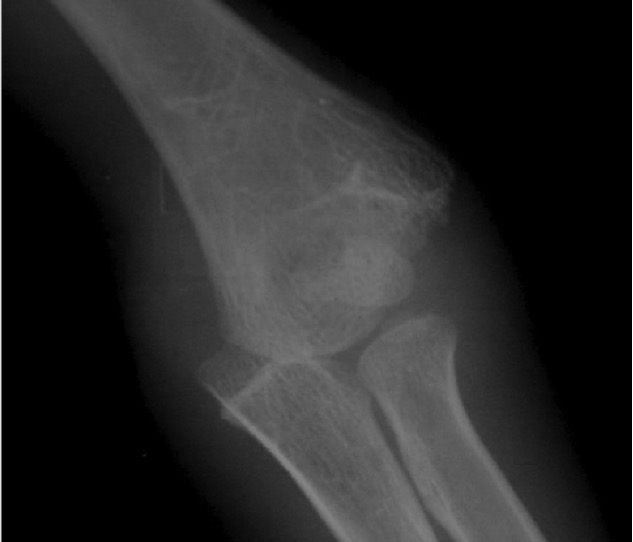
Distal Humeral Physeal Separation
Pathology
Children < 6
- entire distal humerus physis is displaced
Xray
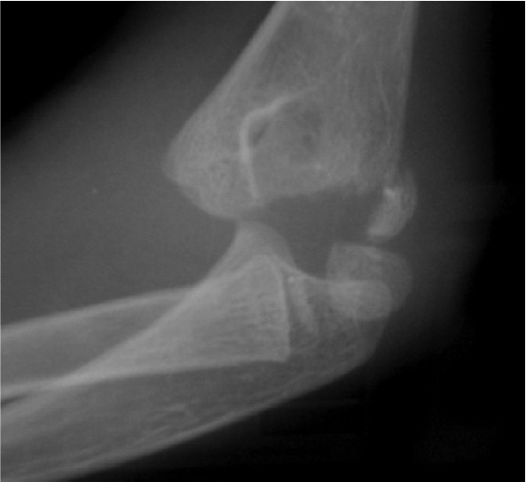
Distal physis not ossified < 1 year
- may be a difficult diagnosis
Children < 6
- entire distal humerus physis is displaced
Distal physis not ossified < 1 year
- may be a difficult diagnosis
> 10 mg / dl
- must be corrected for albumin
Malignancy
- multiple myeloma / lung cancer / breast cancer
Hyperparathyroidism
- elevated PTH
High mortality associated with hypercalcaemia of malignancy
40% albumin bound
50% ionised and active
Fall in level promotes tetanus
Chvostek sign
- tapping masseter muscle induces spasm
Trousseau Sign
- flexion of thumb & wrist with extension of fingers
Carpopedal Spasm
Prolonged QT interval on ECG
1. Vit D Deficiency
Aims of treatment
1. Correct the deformity early
2. Correct it fully
3. Hold the corrected position until foot stops growing
- AFO
- Denis Browne Boots
Timing
Start 1 - 3 weeks
- let parents settle and get used to diagnosis
- explain method and length of treatment required
Decreasing incidence in recent decades most likely attributable to preoperative antibiotics
Conventional discectomy </= 1%
Fusion 2%
Fusion & instrumentation 5-6%
Instrumentation doubles infection rate in lumbar fusion
Diabetes
Most common pattern cord injury
Hyper-extension injury in middle aged man with osteoarthritic spine
Usually C3/4 and C4/5
Most common type / in older patient with pre-existing spondylosis / OPLL
- hyperextension injury
- compression of the cord
- anteriorly by osteophytes
- posteriorly by infolded ligamentum flavum
Bilateral Pars Fracture C2
- traumatic axis spondylolisthesis
Neurological injury uncommon
- fragments separate and decompress
Different to judicial hanging where spinal cord is severed
Rare
- unilateral
- bilateral
Compression
Lateral Compression
Rotation
Skull base pain
Cock Robin
Cranial nerve injury
Type I
Impaction of a condyle
Facet joint dislocations secondary flexion distraction injury
10%
1. Unifacet subluxation - interspinous process widening
2. Unifacet dislocation - 25% anterolisthesis
3. Bifacet dislocation - 50% anterolisthesis
4. Complete vertebral translation - 100% anterolisthesis
Disseminated Intravascular Coagulation
Results from excessive activation of either extrinsic or intrinsic coagulation pathway
- multiple small clots
- consumptive coagulopathy
1. Excessive Extrinsic Activation
Secondary to extensive cellular destruction
- thromboplastins +++ released into circulation