
Epidemiology
NF1 / Von Recklinghausen
- most frequent of these disorders
- 1/3000 - 1/4000.
NF2
- VIII nerve schwannomas
- nil musculoskeletal manifestations
Aetiology
AD
- 50% due to new mutation
Gene protein neurofibromin
Diagnosis
Young children
- multiple café-au-lait spots that appear in the first year of life.
The NIH Consensus Development Conference Statement
- diagnostic criteria for NF1 are met in an individual who has two or more of the following:
1. Six or more café-au-lait spots over 5mm in prepubertal individuals and over 15mm in postpubertal individuals
2. Two or more neurofibromas of any type or one plexiform neurofibroma
3. Freckling in the axillary or inguinal region
4. Optic glioma
5. Two or more Lisch nodules (iris hamartomas)
6. A distinctive osseous lesion such as sphenoid dysplasia or thinning of the long bone cortex with or without pseudarthrosis
7. A first degree relative (parent, sibling, or offspring) with NF1 by the above criteria.
Clinical Features
Café au lait
< 2 normal
6 or more > 0.5cm, smooth edges
Aid early suspicion of NF1
Neurofibroma
Cutaneous
- raised over the skin
- bluish in colour
- increase in number ofter puberty and with pregnancy
- nil malignant potential
- develop from small cutaneous nerves
Plexifom
- bag of worms feeling
- usually develop from major nerves
- potential to become malignant
Axilllary / Inguinal Freckling
Aids early diagnosis
- second most common feature in children
- 80%
Lisch Nodules
Hamartomas in iris
- 90% patients over 6
Optic Glioma
&0% found in NF1
- usually benign and small
- can sometimes rapidly increase in size
Elephantiasis
Dermatological manifestation
- large soft tissue masses
- rough, raised vilous skin
Verrucous Hyperplasia
- skin overgrowth
- velvety, soft papillary
Issues
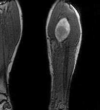
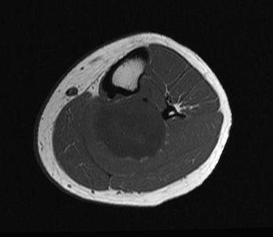
Malignancy
- 30 - 50 years
- large deep lesion
- increasing in size
- apin
Congenital Tibial Pseudoarthosis
- normally 1 per 140 000
- 3% of NF1
- see article
Spinal Deformities / Kyphosis / Scoliosis
- 30% have spinal deformity
- dystrophic and non dystrophic
- recommend earlier fusion at curves > 35o
- higher incidence of pseudoarthrosis
- all require MRI prior as high incidence of intraspinal lesions
- see Paediatrics / Spine / Scoliosis / Other / Neurofibromatosis
Spine
Epidemiology
2% scoliosis patients have NF
10 - 30 % NF patients have spinal deformity
- most common skeletal manifestation
- mostly non dystrophic variety
Types
A. Non dystrophic
- vertebral wedging, angulation and rotation
- very similar to idiopathic scoliosis
- right thoracic
B. Dystrophic
Cause
- may be due to intraspinal lesions such as tumours, meningoceles and dural ectasia
- can simply be bony dysplasia
Characterised by
- short segments, sharp angulation with severe apical rotation
- scalloping posterior margins
- widening of the spinal canal
- enlargement of neural foramina
- widened interpedicular distance
- thinned defective pedicles and lamina
- paraspinal mass
- rotation of ribs - 'pencilling'
Other problems
Cervical spine kyphosis
Atlantoaxial dislocation
- has been reported in five patients
- flexion and extension views are important in assessing cervical instability
- needed pre-op
Kyphosis
- marked acute posterior angulation
- can develop myelopathy and even paralysis
Intraspinal Tumours
Dural Ectasia
- circumferential dilatation of dural sac
- contains CSF and brown material
- erodes osseous structures
- very thin lamina
- can cause great angular deformity
- may have multiple dumbbell appearances
Dumbbell lesion
- single neurofibroma
Meningocoeles / pseudomeningocoeles
Brace treatment
Not been effective in dysplastic curves
Can use early in non dysplastic
Operative Management
Non dystrophic
Issues
Recommend posterior fusion for progressive
- lower threshold for surgery than in idiopathic
- potential for progression much higher
Surgery as for idiopathic
Idiopathic
Brace 20 - 35o
Surgery > 35o
Dystrophic
Issues
A. All dystrophic curves need MRI to assess intraspinal lesion
B. All need C spine screening before surgery
- these patients also often have cervical spine abnormalities and instability
C. Incidence of pseudoarthroses ranges from 6% to 38% with isolated posterior fusion
- recomment addition of anterior fusion for dysplastic scoliosis is therefore recommended
- anterior release then posterior fusion
Indications
Anterior release and fusion followed by posterior fusion if > 50o
Kyphoscoliosis
Posterior fusion alone is usually insufficient in patients with kyphosis
Decompression of the cord with laminectomy
- contraindicated because the lesion is usually anterior
- removal of the posterior elements predisposes the patient to further postlaminectomy kyphosis.
- need decompression and fusion
> 50o
- brace early
- need anterior and posterior fusion
- may even need brace post surgery
- if have myelopathy symptoms may also need decompression
Lordoscoliosis
- in dystrophic