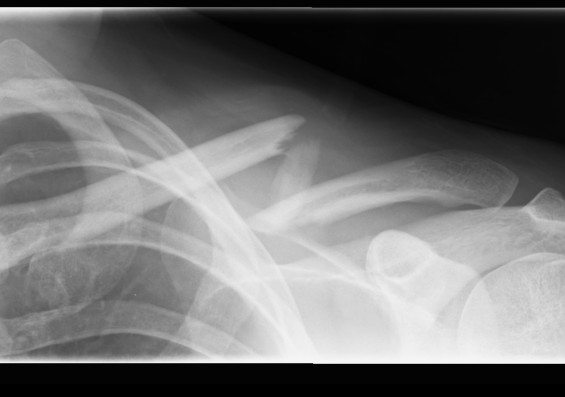
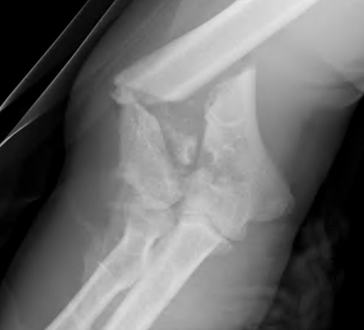
complications

Complications

Intraoperative glenoid fracture


Avoid by

- careful reaming and drilling osteoporotic bone


Management

1.  Rotate metaglene

- use locking screws to stabilise glenoid

2.  PA screws

- cannulated 4.0 mm screws

- inserted percutaneously from posterior


Haematomas


Great deal of dead space is created

- always use a drain

Uncemented femur

GoalTHR Uncemented


Initial press fit

- implant geometry fits the cortical bone in the proximal femur

- good initial mechanical stability


Biological fixation for success

- good press fit

- minimal micromotion

- bony or fibrous tissue ingrowth or ongrowth


Clavicle Fractures

Clavicle Fracture Displaced

Mechanism


Usually a direct blow 

- less commonly a fall on the outstretched hand


RTA / sporting accidents commonest causes


Can be pathological as a result of radionecrosis

- eg following radiotherapy for breast cancer.  


Incidence


Fractures of the clavicle are common

Distal Humerus Fractures

EpidemiologyDistal Humeral Fracture


2 groups

- young patient with high velocity injury

- older patient with comminuted, osteoporotic fracture


In the second group fixation can be very difficult


Anatomy


Hinged Joint

- trochlea axis is centre of rotation

- 40o anterior angulation in sagittal plane

Arthroplasty

Indications


RA 

- very good results

- 97% 10 year survival Coonrad-Morrey prosthesis


Other Dx 

- OA / post-traumatic arthritis / nonunion

- tend to have worse survival than RA


Haemophilia

- elbow joint commonly involved

- 90% of haemophiliacs


Acute unreconstructable fracture > 60