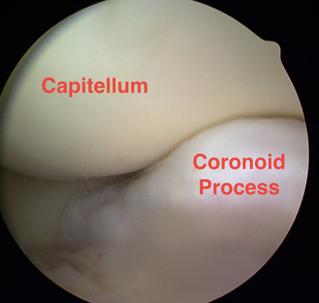
Indications
| Capitellar OCD | Early elbow osteoarthritis and stiffness | Synovectomy / Washout | Tennis elbow |
|---|---|---|---|
|
Removal of loose bodies Microfracture |
Removal loose bodies Excision of osteophytes Release anterior capsular contractures
|
Rheumatoid arthritis Sepsis |
|
 |
 |
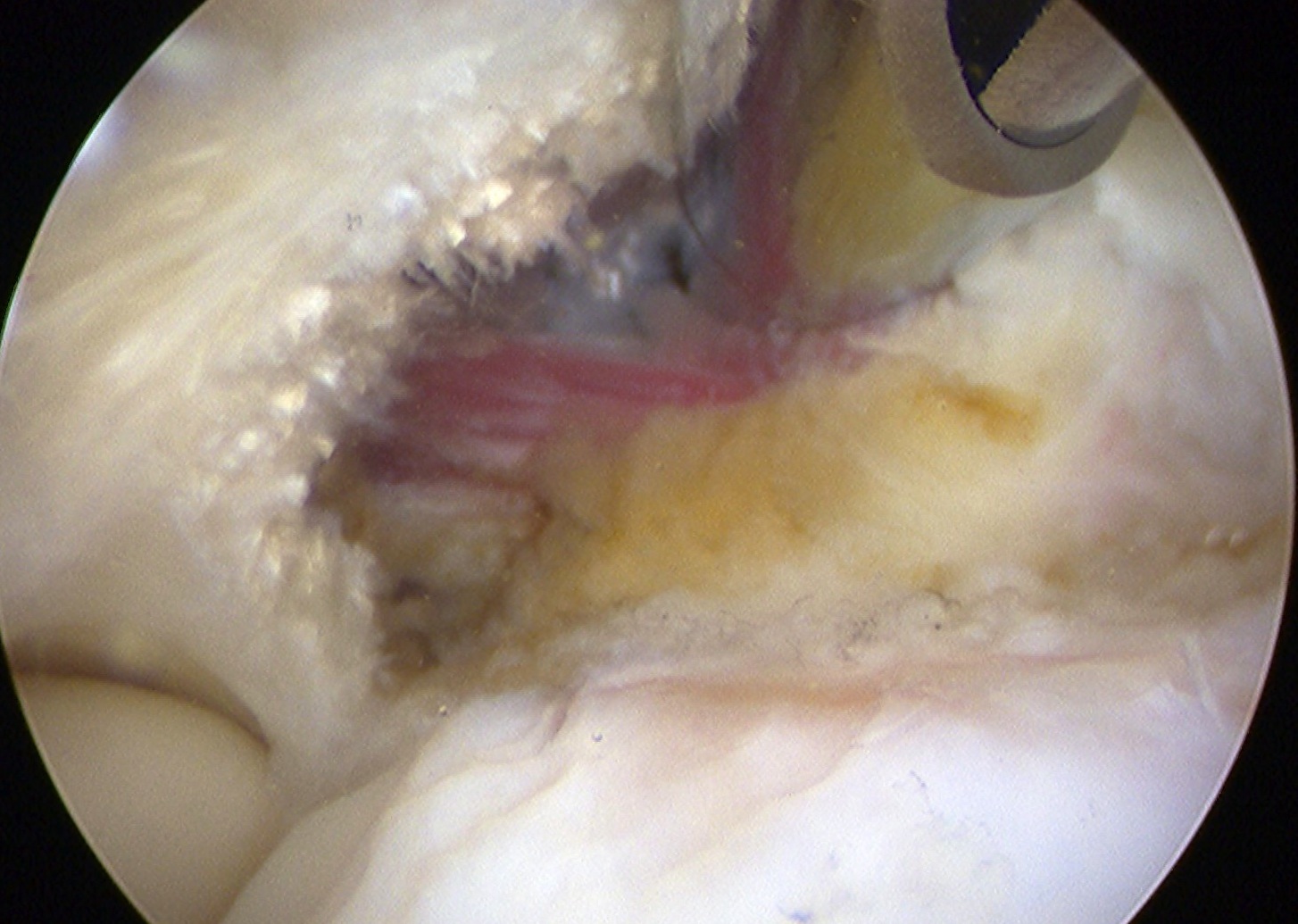 |
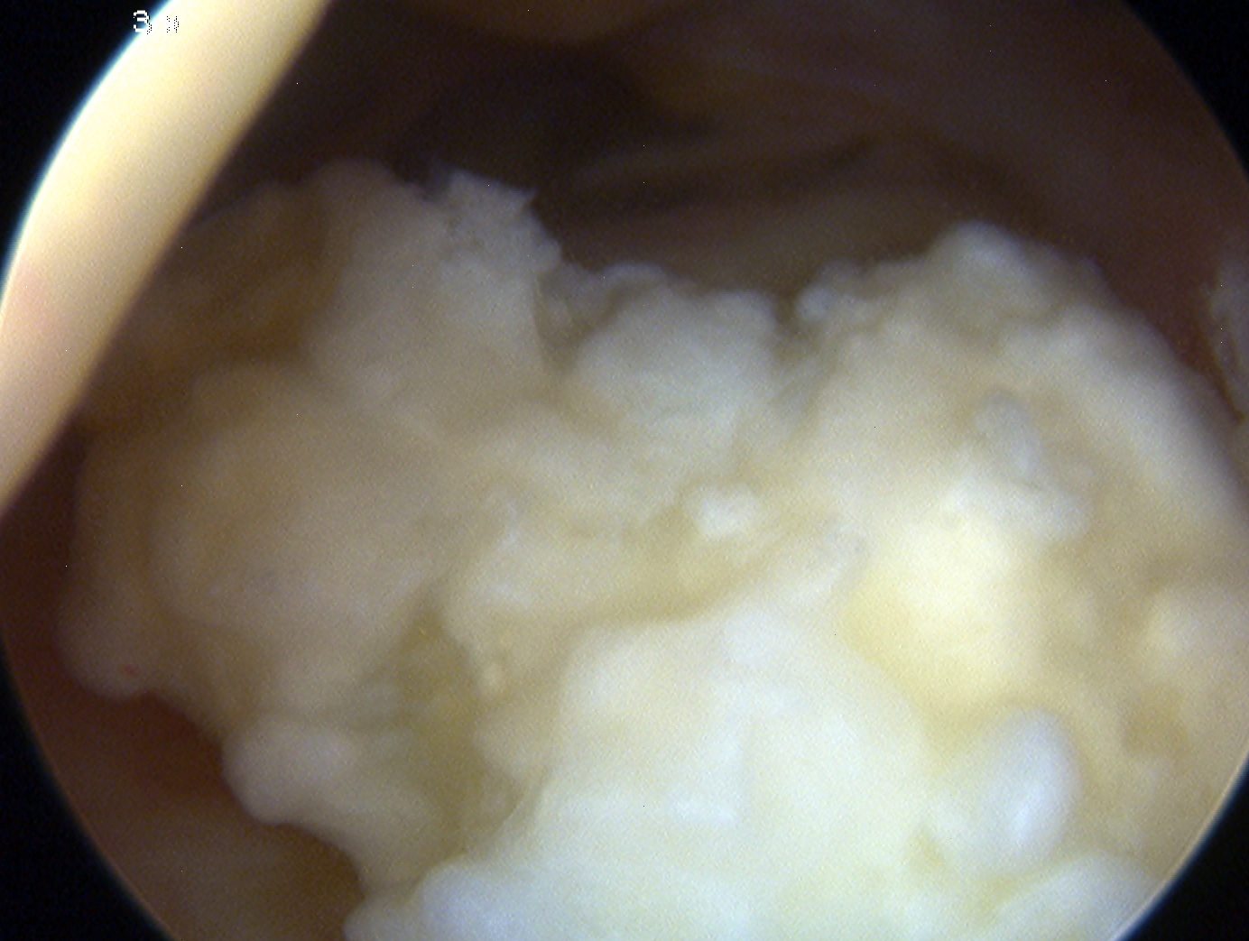
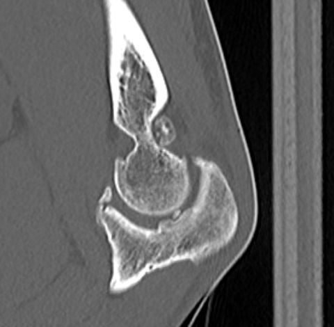
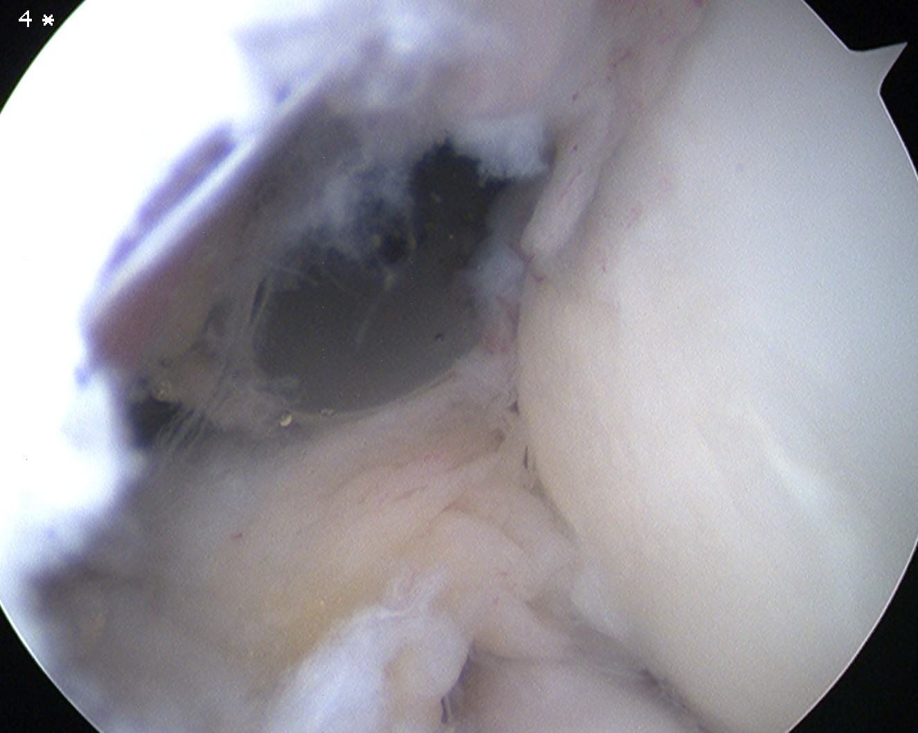
Multiple elbow loose bodies
Single loose body in adolescent
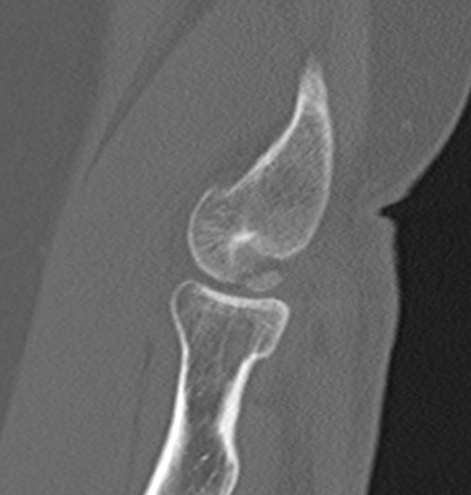
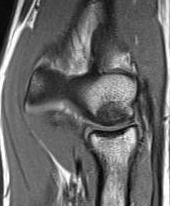
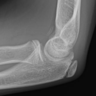
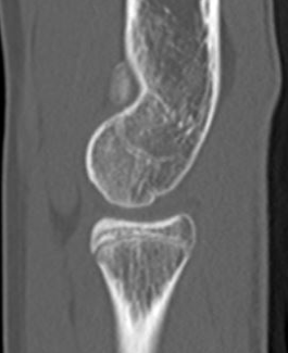
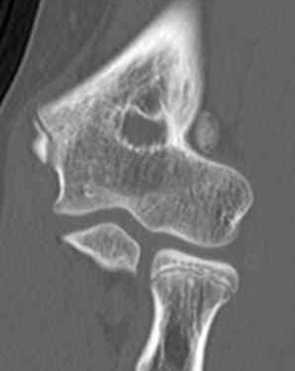
Capitellar OCD www.boneschool.com/capitellar-OCD
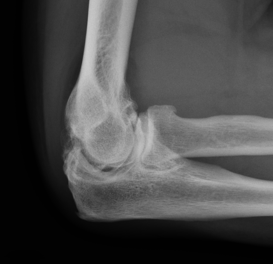
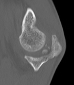
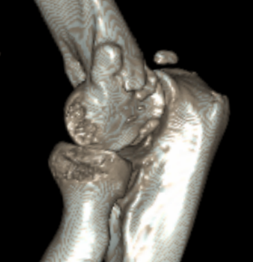
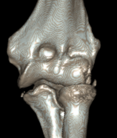
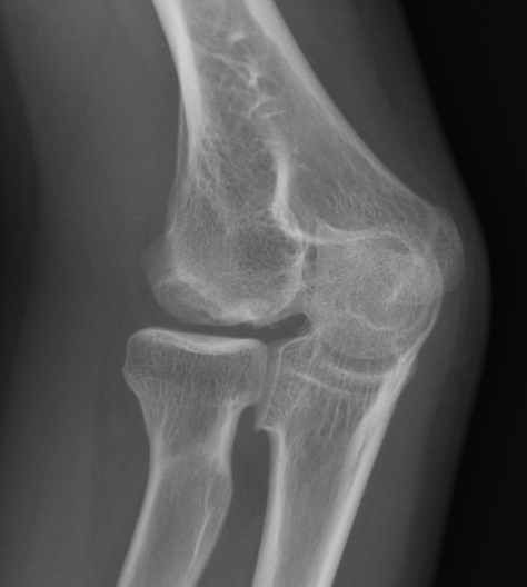
Elbow osteoarthritis and & stiffness www.boneschool.com/elbow-OA
Relative contra-indications
Abnormal elbow scarring
Extensive heterotopic ossification
Previous ulna nerve transposition
Ulna nerve subluxation
Complications
Intravia et al Arthroscopy 2020
- 560 consecutive elbow arthroscopy cases
- 3.5% transient nerve palsy (8 ulnar, 8 radial, 1 median, 3 medial antebrachial cutaneous)
- 2.5% heterotopic ossification
- 0.5% deep infection
- systematic review of 95 studies and 14,000 elbow arthroscopy cases
- overall complication rate 11%
- 4.5% postoperative stiffness
- 4% revision surgery
- 3% nerve injury - ulna nerve most commonly injured
Elbow arthroscopy technique
Vumedi elbow arthroscopy video 1
Vumedi elbow arthroscopy video 2
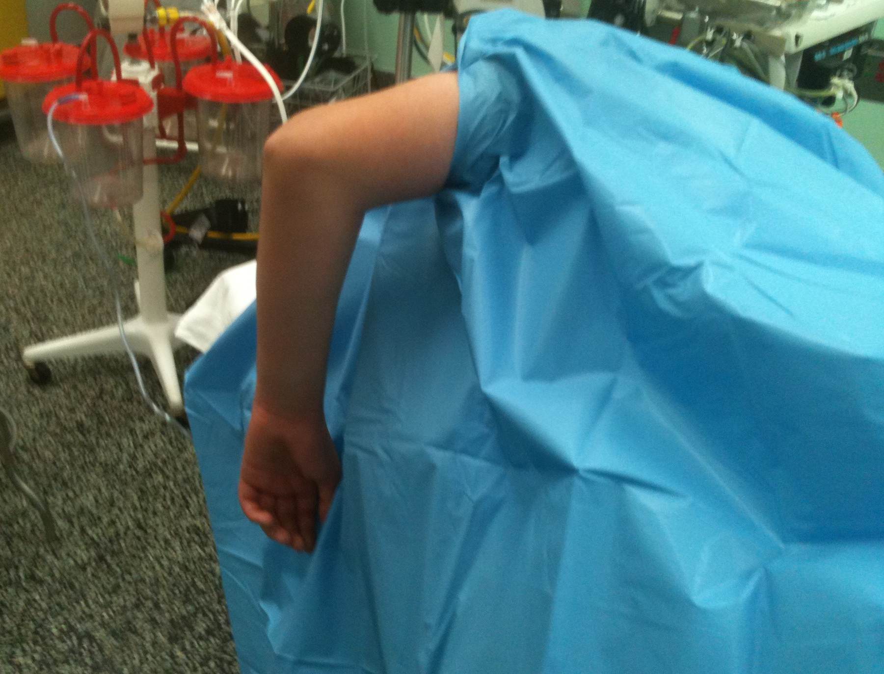
Position
Lateral decubitus
- arm over L shaped bolster
- tourniquet to 250 mmHg
Mark
- medial and lateral epicondyles
- radial head
- olecranon
- ulna nerve
Soft spot
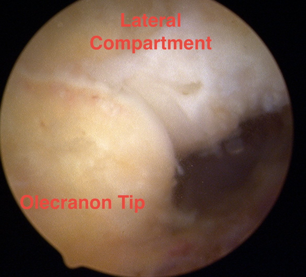
- between lateral epicondyle and olecranon and radial head
- insufflate joint with 30 mls of saline through soft spot
- standard 4mm arthroscopy instrumentations
Portals
| Anterior elbow arthroscopy | Posterior elbow arthroscopy |
|---|---|
|
Proximal anteromedial portal - viewing portal |
Posterocentral - viewing portal |
|
Proximal anterolateral portal - working portal |
Posterolateral portal - working portal |
| Direct lateral portal | Accessory posterolateral portals |
Anterior elbow arthroscopy
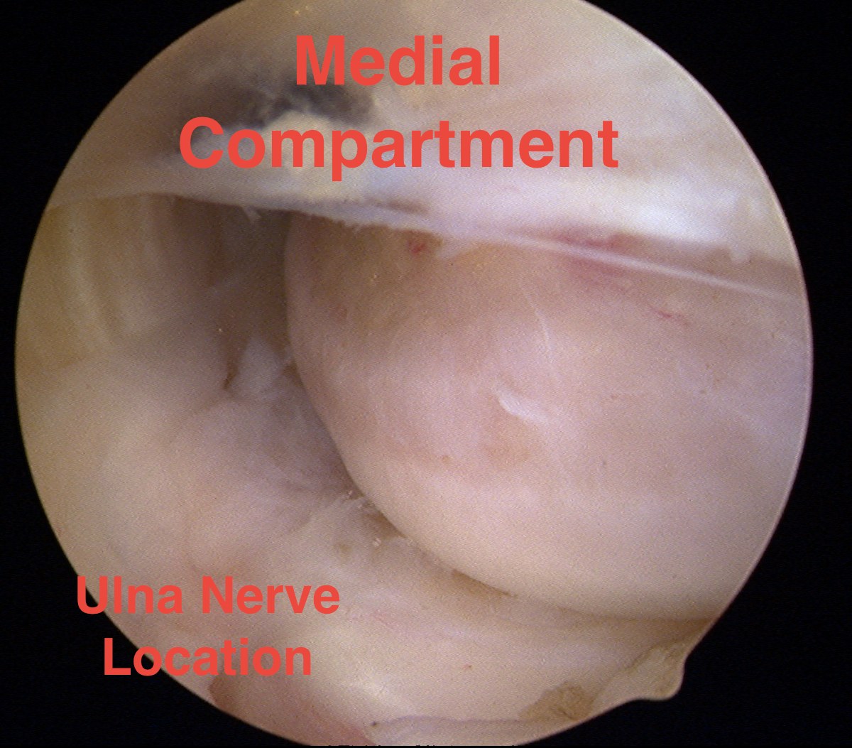
Proximal anteromedial portal
Technique
- viewing portal
- 2cm proximal to the medial epicondyle
- just anterior to humerus / medial intermuscular septum
- blunt dissection and insert portal
Risk
- ulna nerve posterior and behind medial epicondyle
- median nerve and brachial artery anterior
Cushing et al Arthroscopy 2019
- systematic review of safety of anteromedial portals
- proximal AM portal safer than AM portal
- flexion of the elbow improves safety
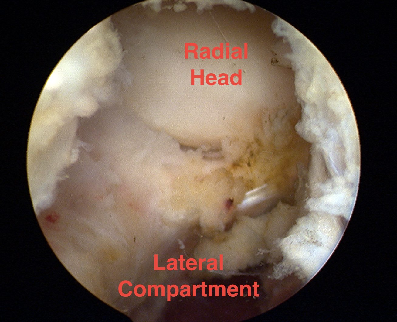
Proximal anterolateral portal
Technique
- 2 cm proximal to lateral epicondyle
- just anterior to lateral intermuscular septum
- outside in technique with needle towards coranoid foss
Risk
- radial nerve at risk with more distal portal
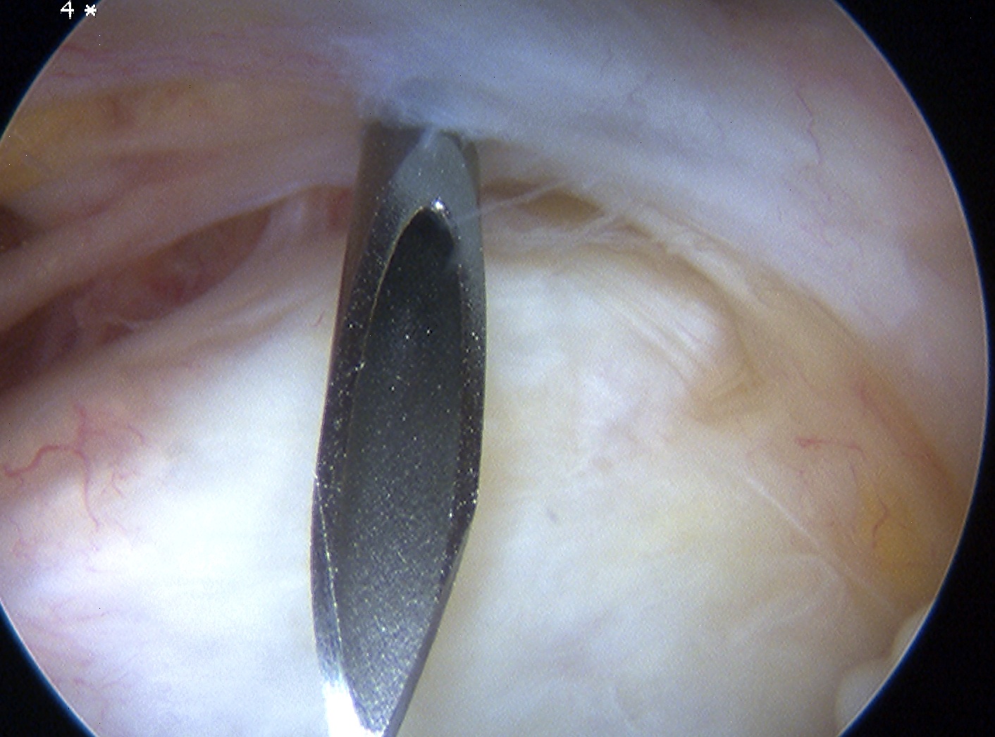
Camera in anteromedial portal creating working anterolateral portal
Direct lateral portal
Technique
- anconeus triangle / soft sport
- olecranon tip / radial head / lateral epicondyle
- through skin, anconeus, capsule
Risk
- posterior cutaneous nerve
Posterior elbow arthroscopy
Indication
Posterior loose bodies
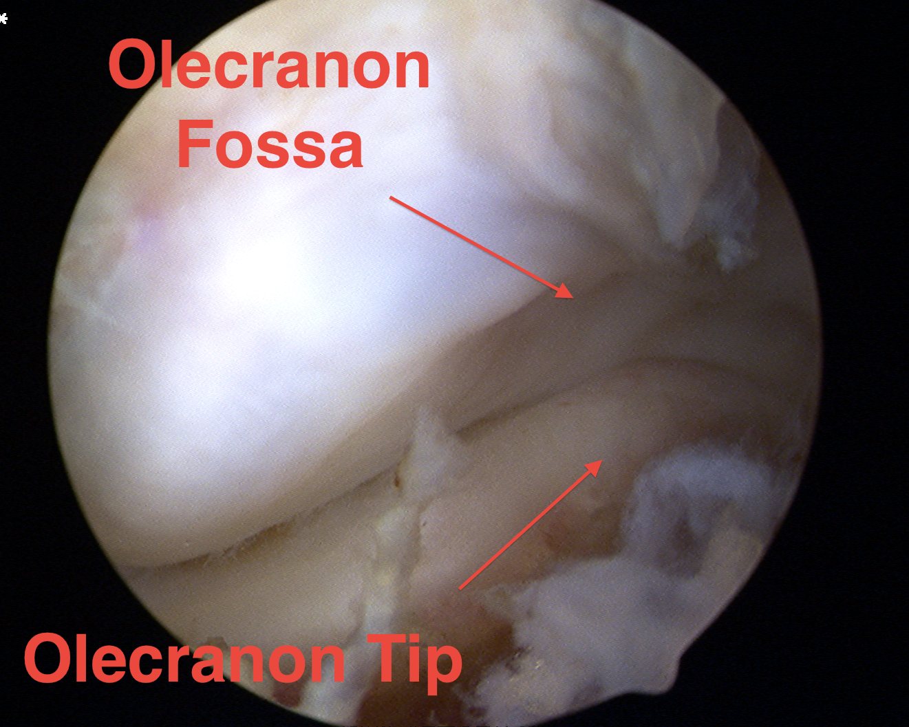
Olecranon tip / fossa impingement
Posterocentral / direct posterior portal
Technique
- viewing portal
- 3 cm proximal to tip olecranon
- in midline through triceps
Risk
- ulna nerve medially
Posterolateral portal
Technique
- 2 - 3 cm proximal to tip olecranon
- in line with lateral edge of triceps
- outside in technique with needle
Accessory porterolateral portals
Technique
- in line with posterolateral portal
- distal as required