Incidence
10 - 15% normal feet
Often bilateral
< 1% symptomatic
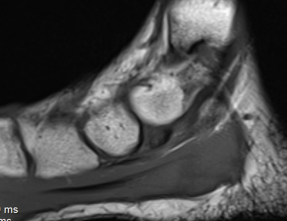
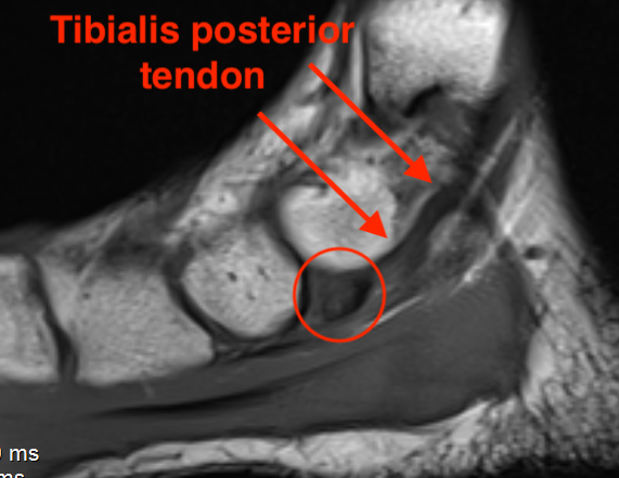
Anatomy
Medial aspect of foot proximal to navicular and part of tibialis posterior tendon
Usually will fuse with navicular
Issues
Pain
- insertional tendonitis / bony prominence / irritation between bone and navicular
- may fracture
Flat foot / pes planus / planovalgus
- accessory navicular probably not a cause of flat foot
- surgical resection / tibialis posterior repair does not resolve pes planus
- some patients may need arthroereisis / osteotomy as well
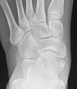
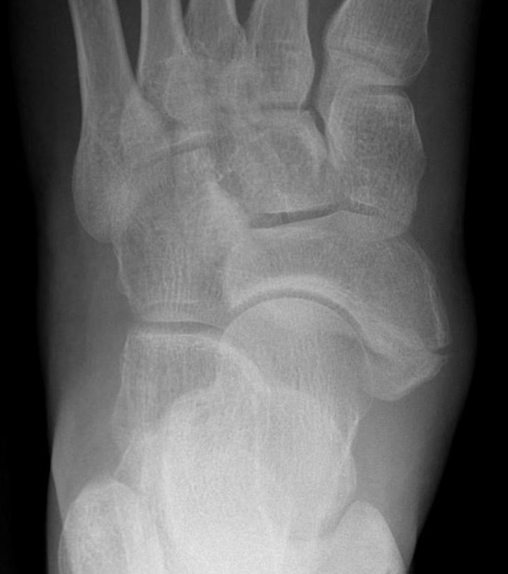
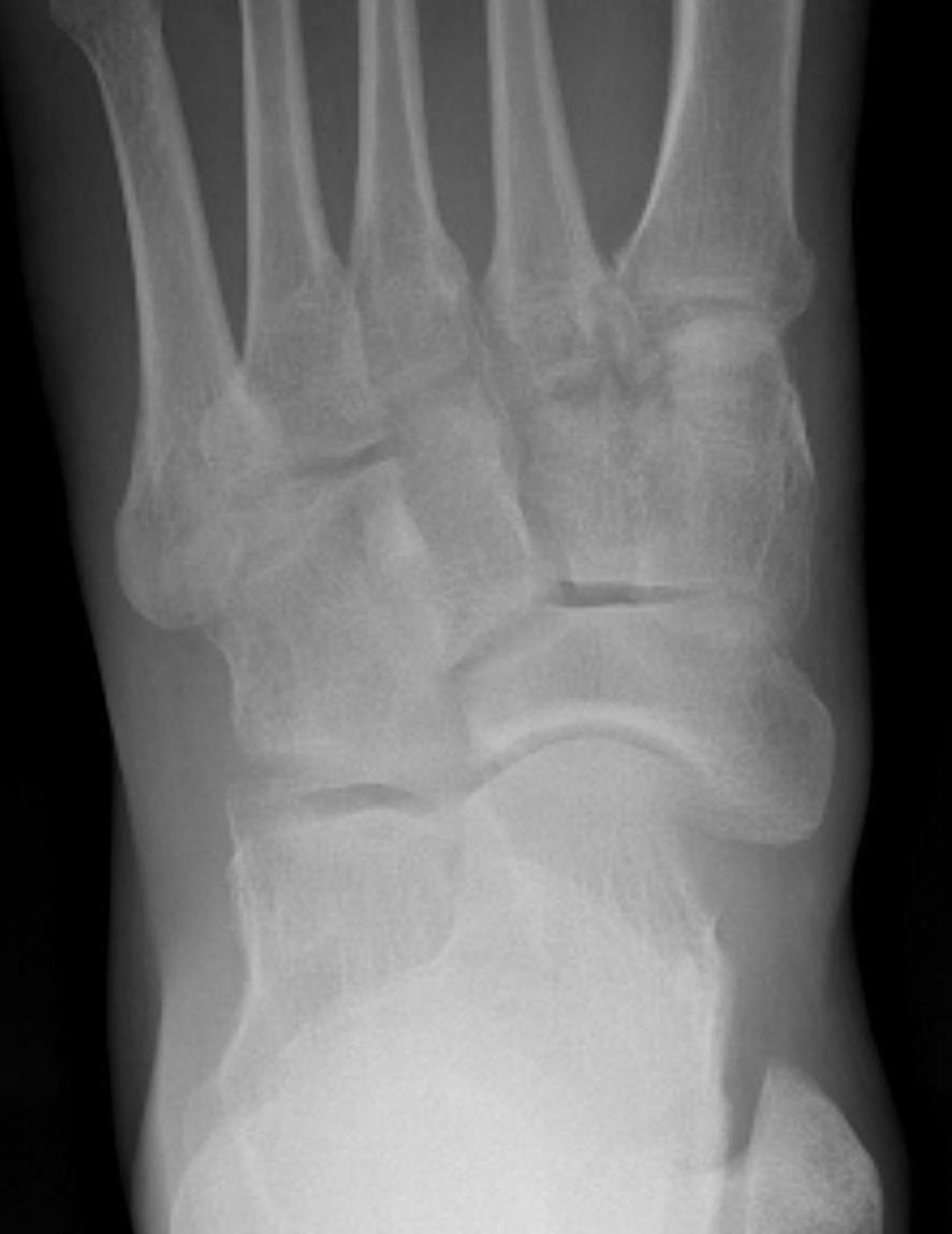
Classification
| Type I | Type II | Type III |
|---|---|---|
|
Small ossicle proximal to insertion In the Tibialis posterior tendon |
Triangular ossicle Connected to navicular via syndesmosis May fracture with injury |
Enlarged medial navicular Cornuate navicular Likely that Type II accessory navicular has fused |
 |
 |
 |
Bone Scan
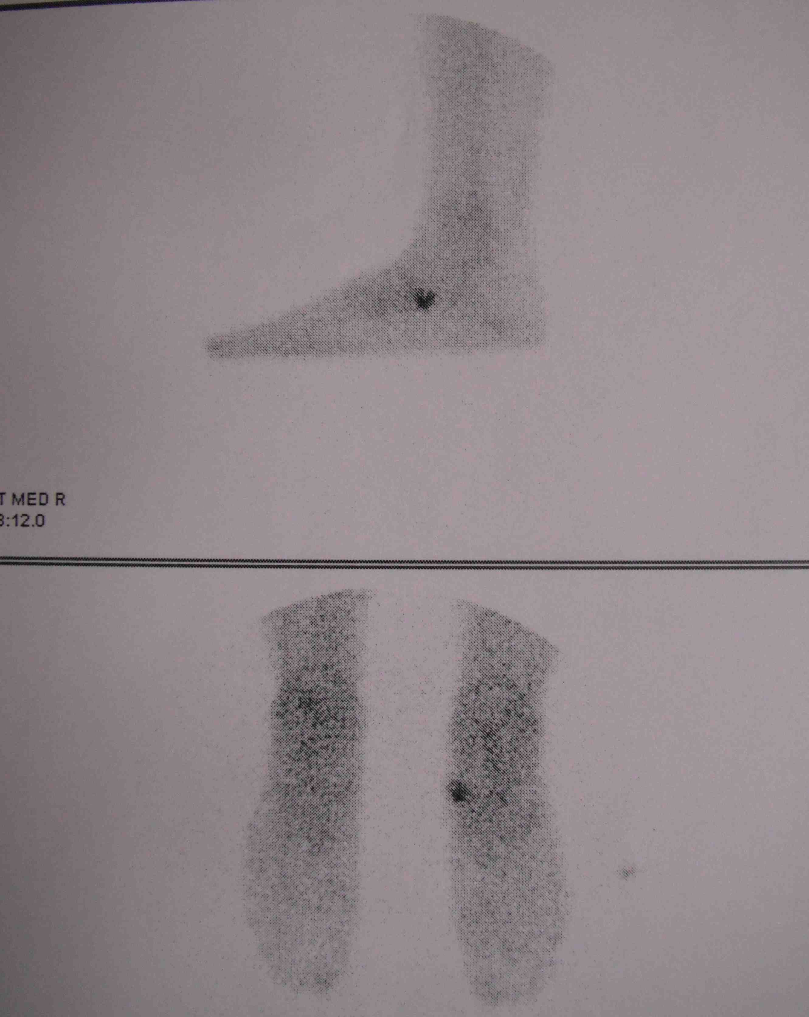
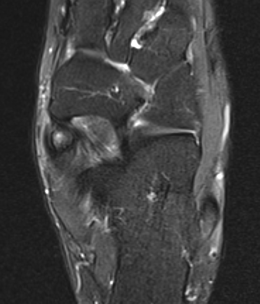
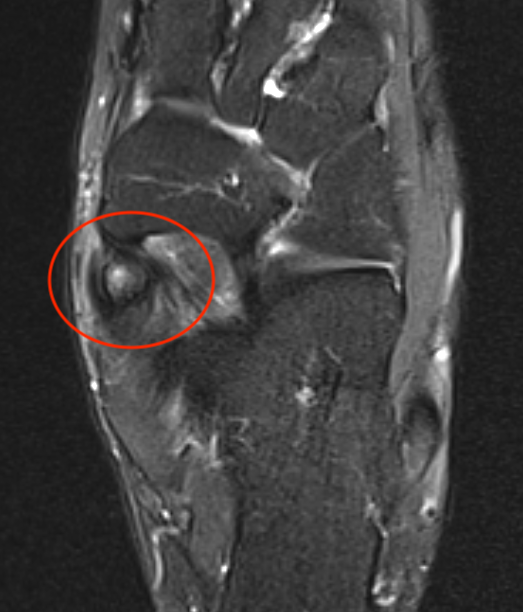
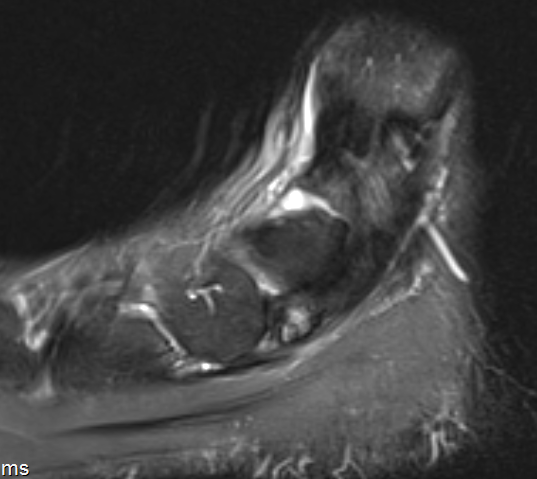
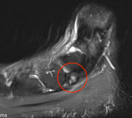
MRI
Show edema about a symptomatic accessory navicular
Differential diagnosis
Tibialis posterior tendonitis
Navicular stress fracture
Nonoperative management
Options
Rest
Walking boot
Orthotics
Cortisone injection
- nonoperative management of 226 symptomatic accessory navicular
- average age 12 years
- Type 2 most frequent (72.7%)
- 28% complete pain relief
- 41% partial pain relief
- 30% required surgical intervention
Operative management
Options
Simple excision
Kidner procedure - excision + tibialis posterior re-routing
Synchondrosis fusion - Type II accessory navicular
Excision / Kidner procedure + subtalar arthroereisis / osteotomy - ? flexible planovalgus foot
Simple excision / Kidner procedure

Simple excision technique
Youtube video endoscopic accessory navicular excision
Kidner procedure technique
Youtube video Kidner procedure
Arthroscopy techniques endooscopic Kidner procedure PDF
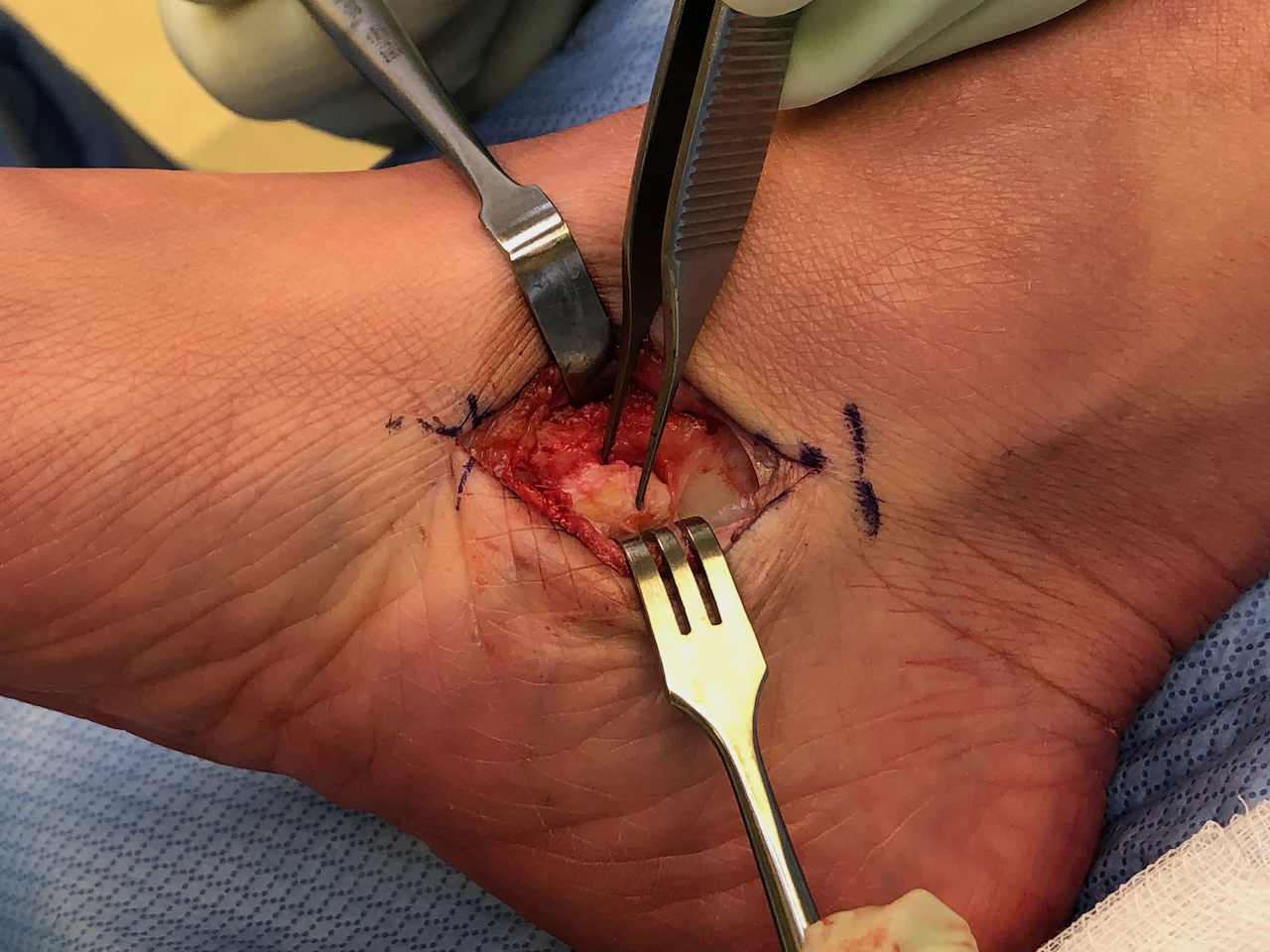
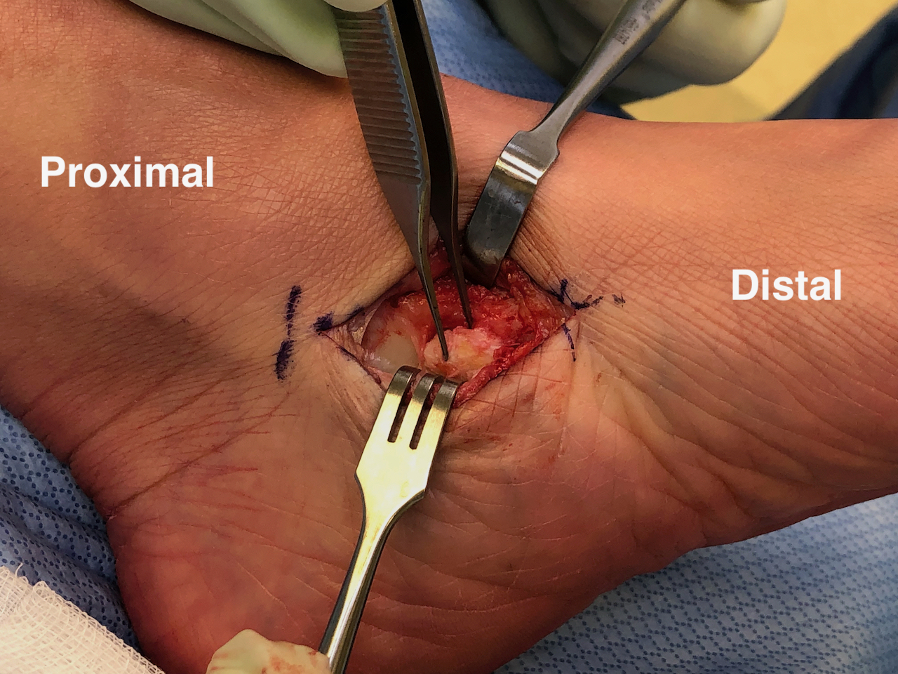
Medial incision dorsally over navicular
- enucleate accessory navicular from tendon
- may need to take away navicular prominence
- reattach tibialis posterior tendon via drill holes / anchor
Results
Wariach et al Curr Rev Musculoskeletal Med 2022
- systematic review of excision versus Kidner
- 164 simple excision: 80% excellent, 18% moderate, 2% poor
- 116 Kidner: 58% excellent, 27% moderate, 13% poor
Accessory navicular / synchondrosis fusion
Indication
Type II
Technique
Arthroscopy techniques endoscopic accessory navicular synchondrosis fusion PDF
Accessory navicular - navicular fusion
- accessory navicular must be large enough / Type II
- debride syndesmosis
- fusion with screw
Results
Chung et al Foot Ankle Int 2009
- fusion of accessory navicular in 34 feet
- 27/34 (79%) good or excellent results
- nonunion in 6/34 (18%)
Zeng et al Foot Ankle Surg 2020
- comparision Kidner to fusion in Type II
- arthrodesis: 83% good / excellent
- Kidner: 80% good / excellent
Planovalgus foot + accessory navicular
Results
- 21 patients with recurrent pain after Kidner procedure
- all had valgus hindfoot treated with calcaneal osteotomy
- improved clinical outcomes
Shi et al J Orthop Surg Res 2023
- 50 flexible planovalgus feet with accessory navicular
- treated with subtalar arthroereisis and medial soft tissue reconstruction
- significant improvement in functional outcomes