Definitions
Tendinitis - inflammation of tenosynovium
Tendinopathy - degenerative changes that can can lead to tears
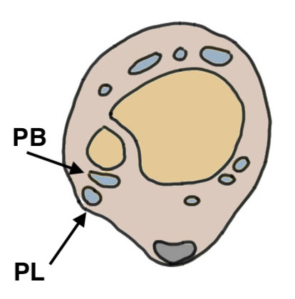
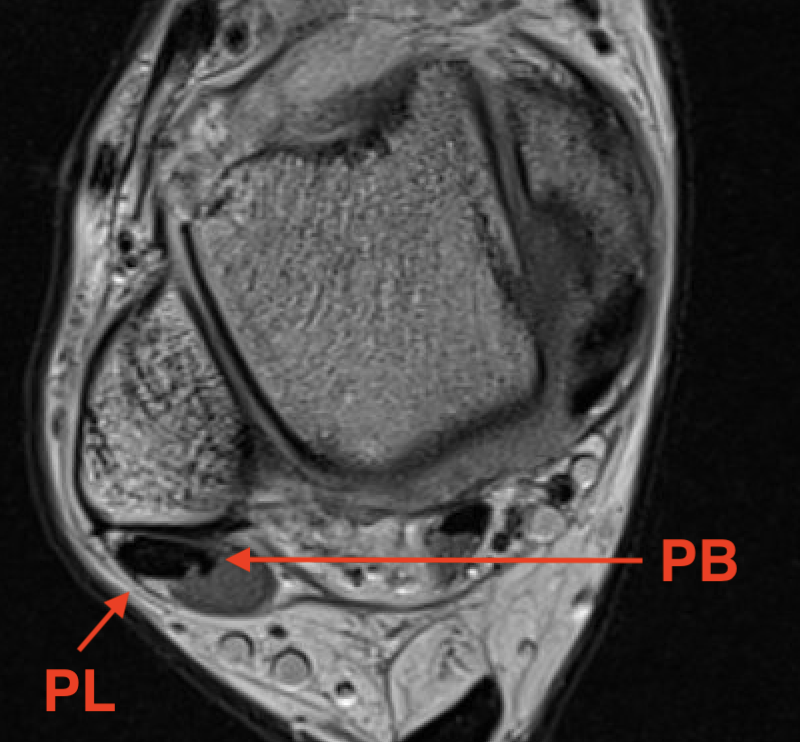
Anatomy
| Peroneus longus | Peroneus brevis | |
|---|---|---|
| Origin |
Lateral condyle of tibia and head fibula Tendon superficial to PB in retromalleolar groove |
Middle third fibula and intermuscular septum |
| Insertion |
Plantar surface base of 1st metatarsal Lateral aspect medial cuneiform |
Tuberosity base 5th metatarsal |
| Action |
Everts the foot Plantar flexes the first ray and ankle Stabilises the medial arch in stance |
Abducts and everts the foot Plantar flexes the ankle |
| Innervation | Superficial peroneal | Superficial peroneal |
Classification
| Zone 1 | Zone 2 |
|---|---|
| Behind lateral malleolus |
Distal to tip of fibula Cuboid tunnel |
| P. brevis more common | P. longus more common |
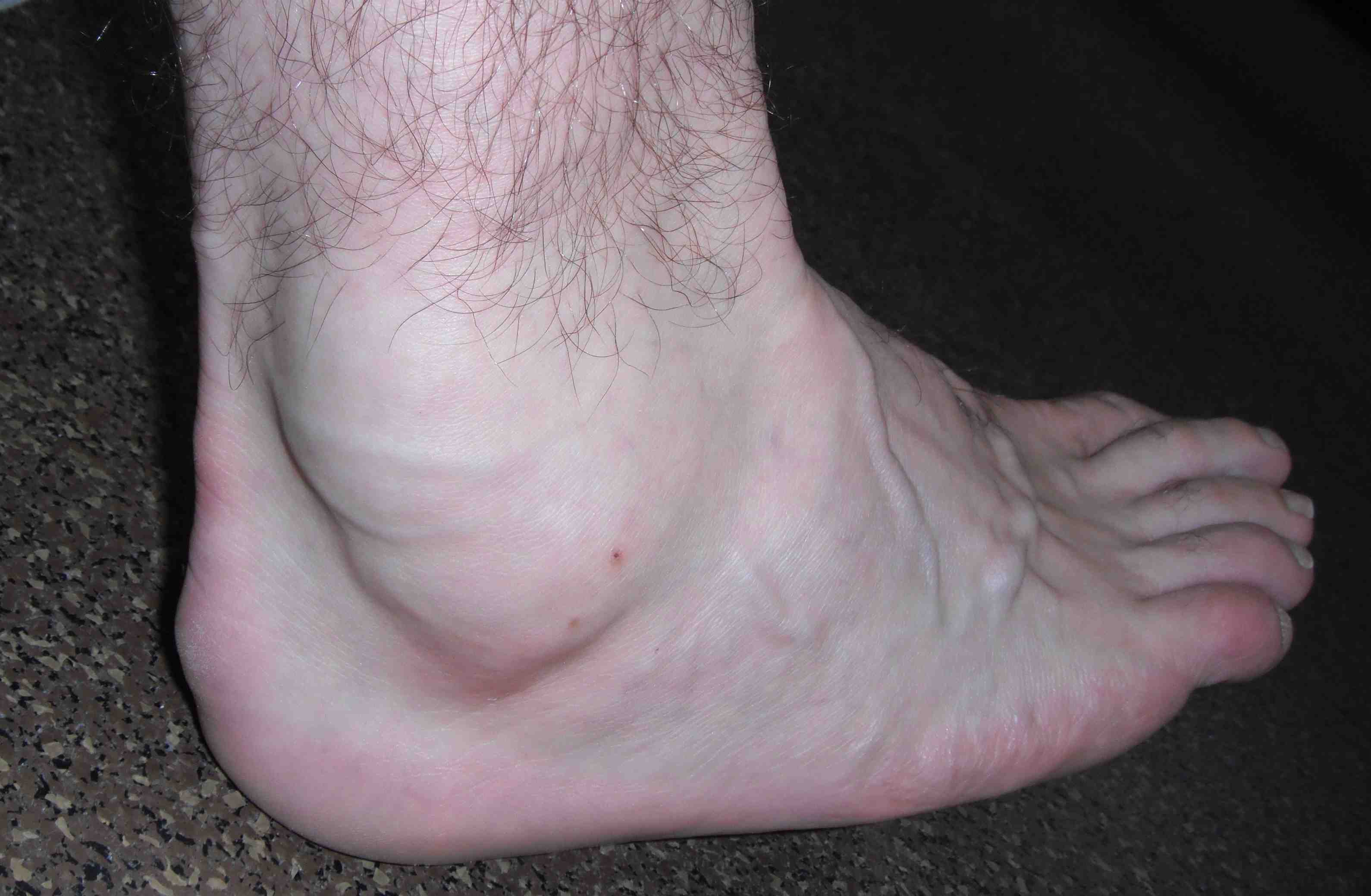
Clinical
Acute or chronic lateral ankle pain
Tenderness / swelling along tendons
Pain with passive inversion and plantarflexion / active eversion
Xray
Usually normal
May see retract os peroneum with P longus tear
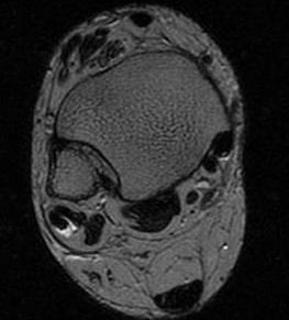
MRI
Findings
- tendonitis - fluid around tendons
- tendinopathy - tendon thickening
- tears
Avoid magic angle effect at ankle - parasagittal obliques / plantar flex ankle
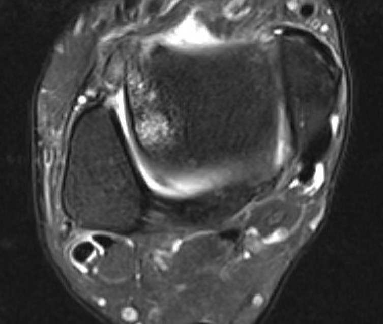
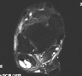
Tendinitis - fluid around tendon
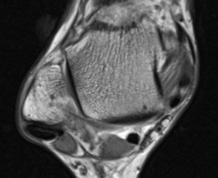
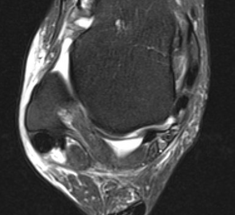
Tendinopathy - thickened PL
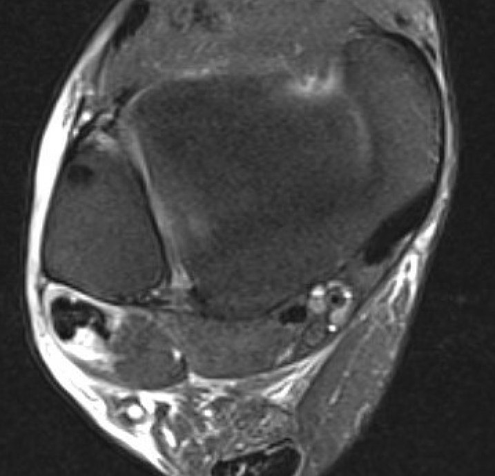
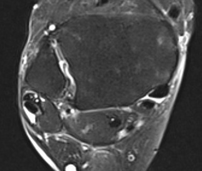
Peroneal brevis tears
Nonoperative management
Analgesia / anti-inflammatories
Modification activities
Lateral heel wedge if hindfoot varus
Boot
Operative management
Options
Tendonitis - debridement and synovectomy
Tendon tears < 50% - debridement and repair with tubularization
Single tendon tears > 50% - tenodesis to other peroneal tendon
Both tendon tears > 50% - tendon transfers / allograft reconstruction
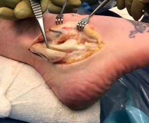
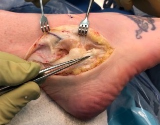
Tendonitis / Tenosynovitis
Open Debridement and tenosynovectomy
Incision along posterolateral border of fibular
- protect sural nerve
- open superior peroneal retinaculum
- synovectomy
Tendinopathy
Endoscopic / Tendoscopy
Vumedi peroneal tendoscopy video
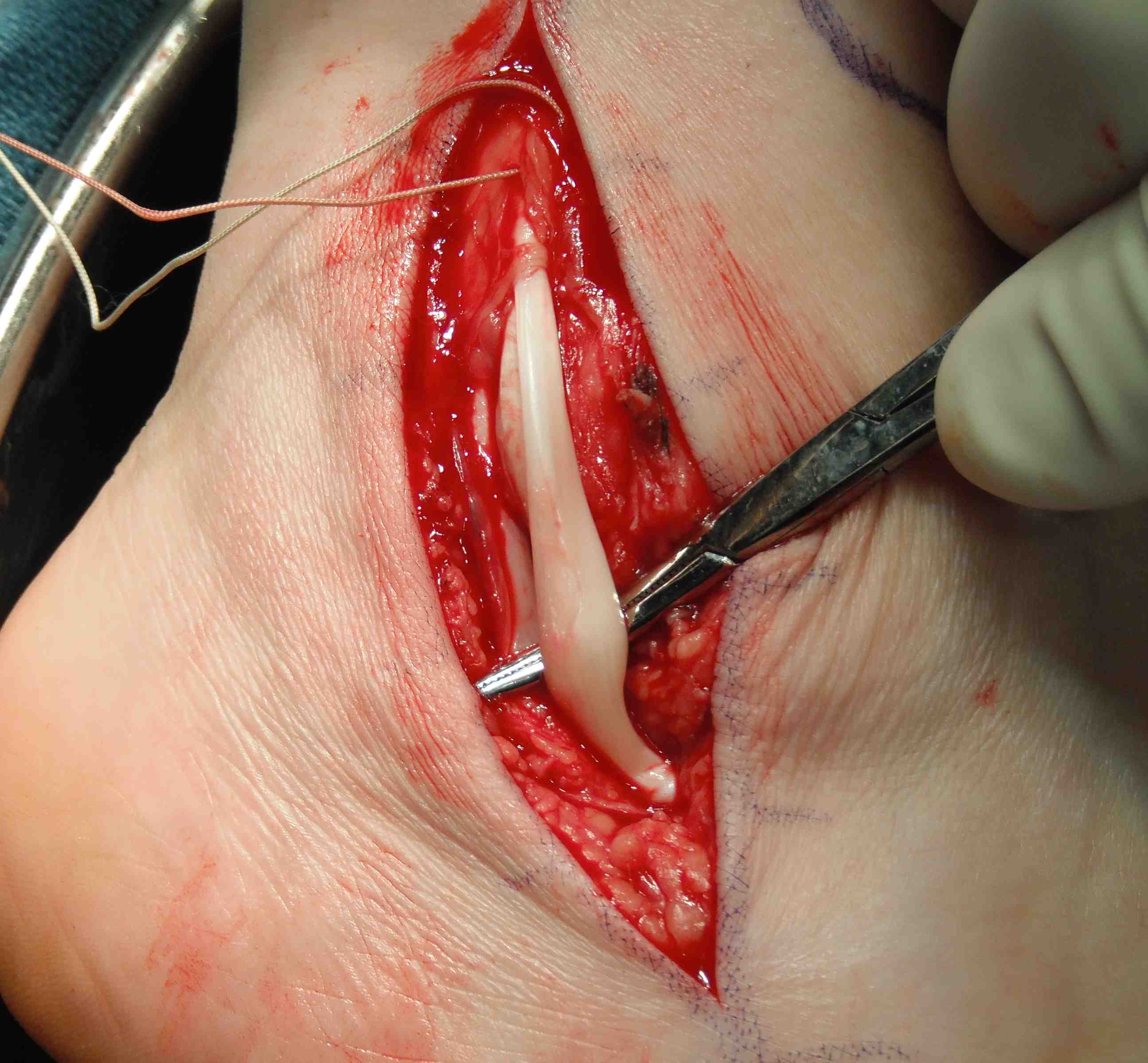
Tendon Tears < 50%
Repair and tubularisation
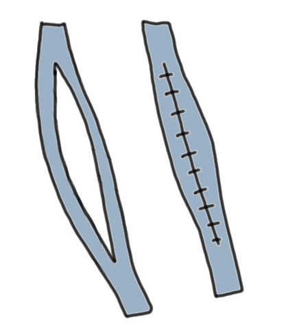
Vumedi P brevis open repair video
Results
Steginsky et al Foot Ankle Int 2016
- 52 patients post primary repair of P brevis
- 86% satisfaction
- 77% return to pre-injury activity at 1 year
Single tendon tears > 50%
Tenodesis
Arthroscopy techniques Peroneal tenodesis PDF
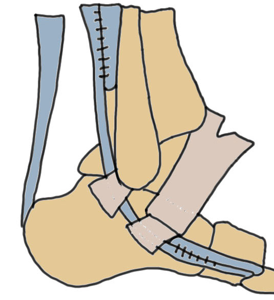
P brevis tears
- 2 part tenodesis
- proximal - P brevis to P longus proximal to superficial peroneal retinaculum
- distal - P brevis to P longus at base 5th metatarsal
P longus tears
- proximal tenodesis to P brevis
Results
Burkhard et al Foot Ankle Int 2021
- 14 patients with P longus to P Brevis transfer
- good clinical outcomes
- no difference in strength with contralateral foot
Both tendon tears > 50%
Lateral transfer of FHL / FDL
Vumedi FHL transfer for peroneal tendon rupture video
Harvest donor tendon at Knot of Henry
- pass posteriorly around tibia / fibular
- tenodesis to tendon proximally or to base of 5th metatarsal
Jockel et al Foot Ankle Int 2013
- 8 patients with FHL / FDL transfer for severe P longus and P brevis pathology
- 7/8 good or excellent results
Allograft reconstruction
Mook et al Foot Ankle Int 2013
- 14 patients undergoing allograft reconstruction
- reductions in pain / improvement in function and strength