Definition
Progressive collapsing foot deformity
Acquired flat foot deformity / planovalgus foot secondary to tibialis posterior dysfunction
Epidemiology
F > 40
Associations - hypertension / diabetes / obesity
Anatomy Tibialis Posterior
| Origin | Insertion | Nerve supply | Action |
|---|---|---|---|
|
Posterior tibia, fibula and inter-osseous membrane
Acute angle around medial malleolus - flexor retinaculum holds tendon in groove - relative hypo-vascular zone 1-2cm distal to medial malleolus |
Navicular tuberosity Plantar cuneiforms 2,3,4 metatarsals Sustentaculum tali |
Tibial nerve (L4/5, S1) |
Plantar flexor ankle joint Inverts subtalar joint Adducts foot Maintains longitudinal arch
Single heel raise - locks the midtarsal joints - allows T Achilles to perform heel raise |
Pathology
Avascular zone
- behind medial malleolus
- area of incomplete mesotenon which provides blood supply
Tendon changes
- paratendinitis - fluid in sheath + synovial proliferation
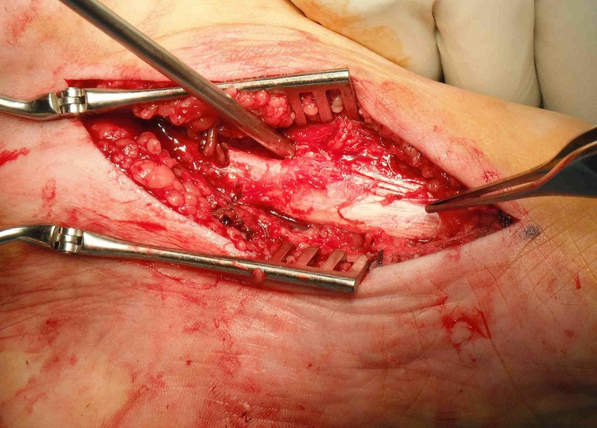
- tendinosis - tendon degeneration with enlargement and longitudinal splits
- elongation of tendon
- rupture
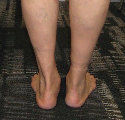
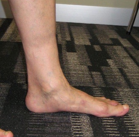
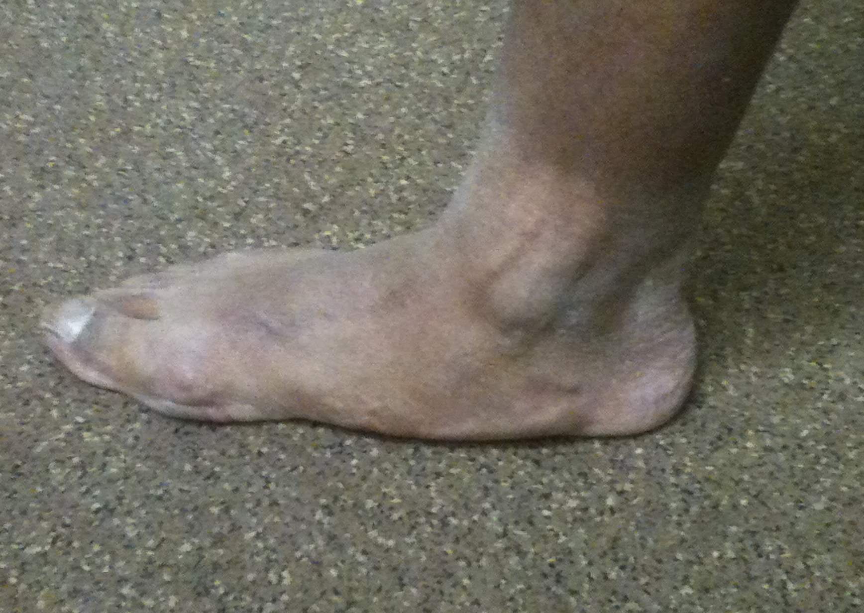
Deformity
Acquired planovalgus
- medial arch collapse
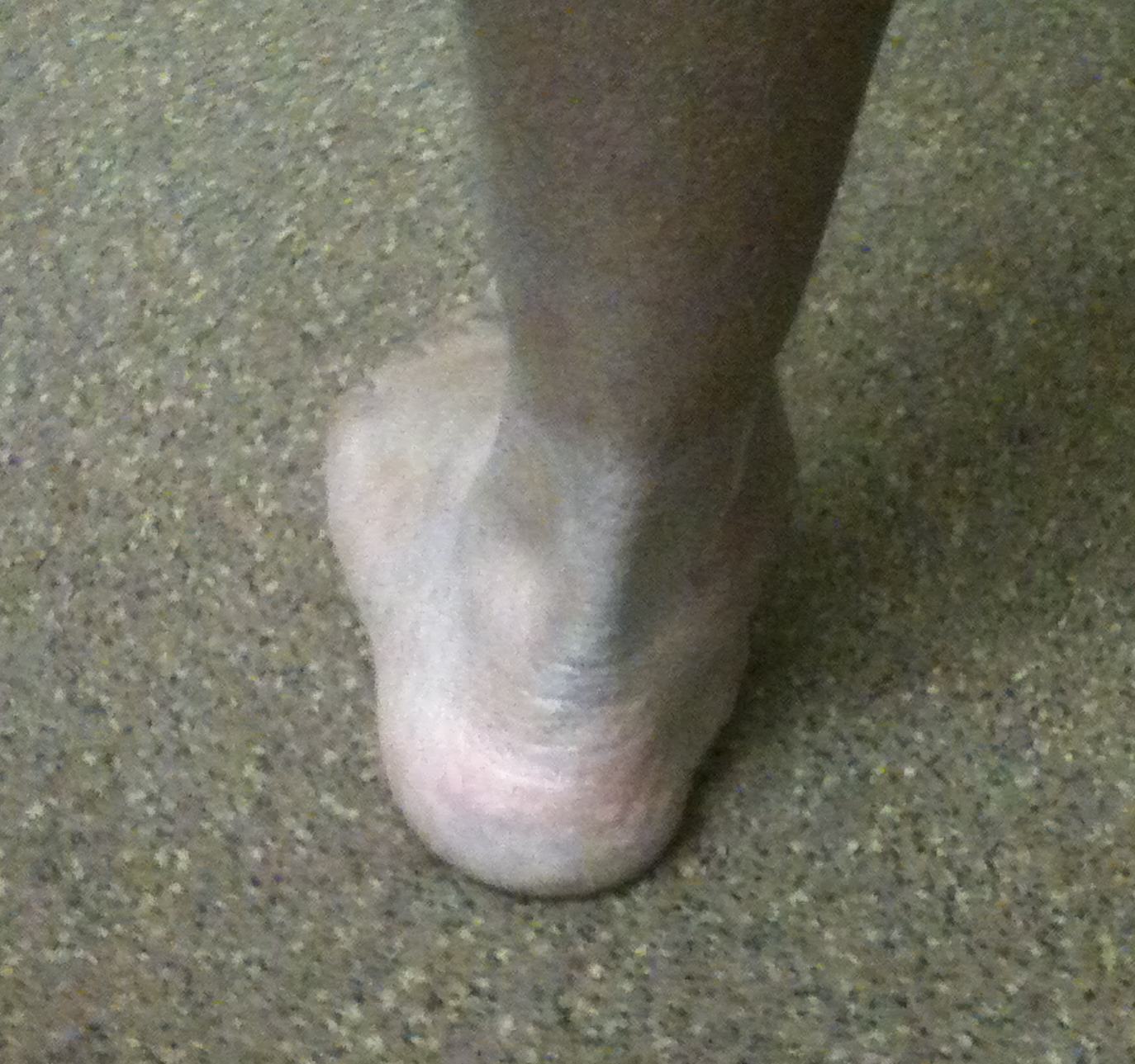
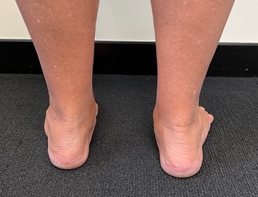
- subtalar joint everts / valgus heel
- foot abducts at TNJ
- Achilles tendon acts as evertor when heel in valgus
- calcaneus impinges on fibular causing lateral ankle joint pain
- attenuation of TNJ capsule, spring ligament and deltoid ligament
Johnson Classification
| Stage 1 | Stage 2 | Stage 3 | Stage 4 |
|---|---|---|---|
| T posterior tendonitis | T posterior elongation / attenuation / rupture | Fixed deformity subtalar joint |
Varus angulation talus in ankle joint |
| Able to single heel raise |
Unable to single heel raise Correctable subtalar joint |
Non correctable valgus | |
|
IIA: As above IIB: Forefoot abduction |
+/- subtalar OA | +/- ankle joint OA |
History
Deformity
Medial pain
Lateral pain due to fibular impingement
Difficulty with shoe wear
Examination
Stage 1
- normal arch with tenderness tibialis posterior tendon
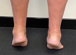
Stage 2
- planovalgus foot - flattened medial arch + valgus heel
- flexible subtalar joint - good ROM, moves into varus with heel raise
- weak tibialis posterior power - foot inverted and in equinus
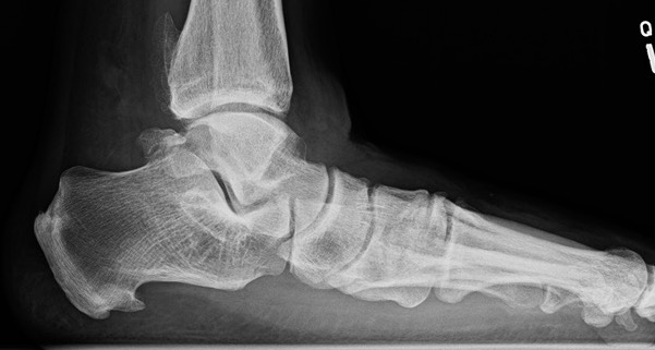
X-ray
Lateral weight bearing
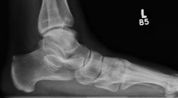
Early - reduced talo-metatarsal angle / Meary's angle
Late - subtalar joint OA
AP weight bearing foot
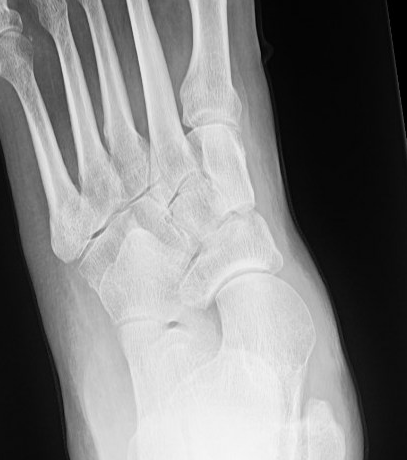
Talonavicular uncovering > 40% - forefoot abduction / stage IIB
AP weight bearing of ankle
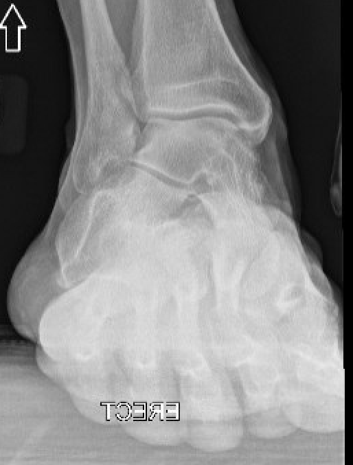
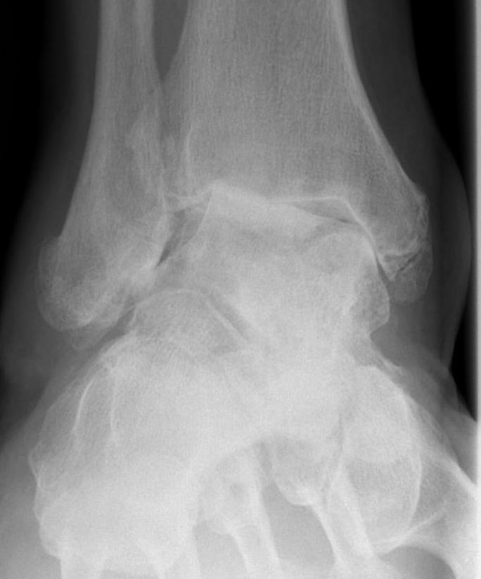
Early - calcaneus under lateral malleolus
Late - valgus tilt of talus with ankle joint osteoarthritis
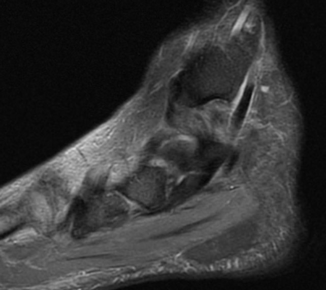
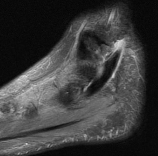
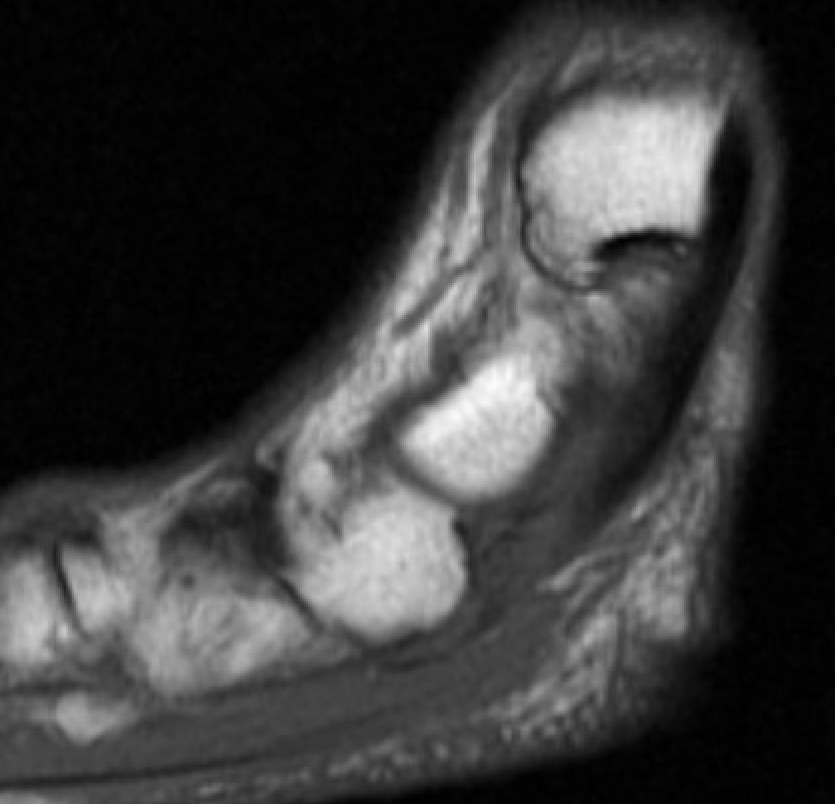
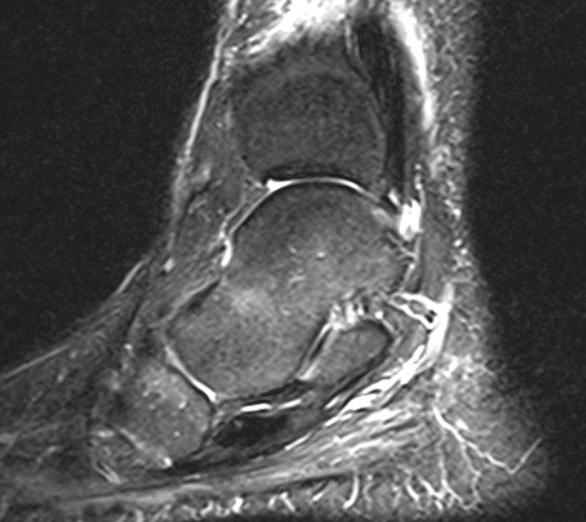
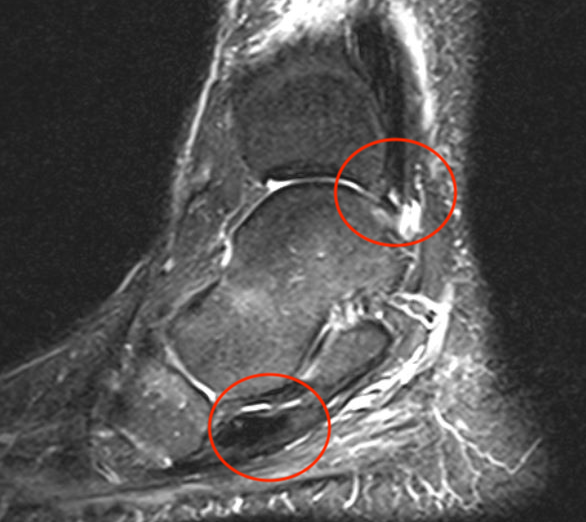
MRI
Tendonitis - fluid around tendon
Tendinopathy - tendon thickening
Tears
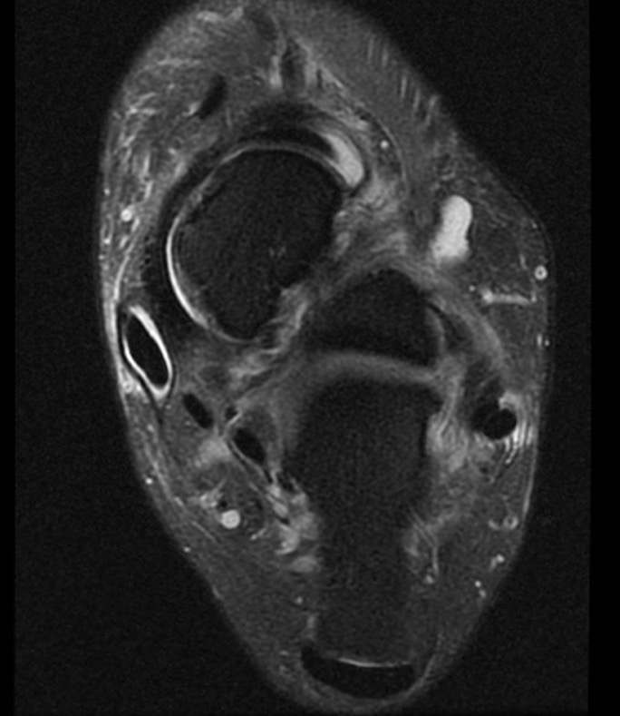
Tibialis posterior tendonitis
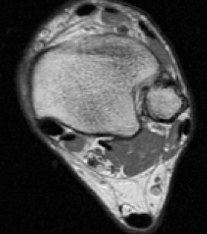
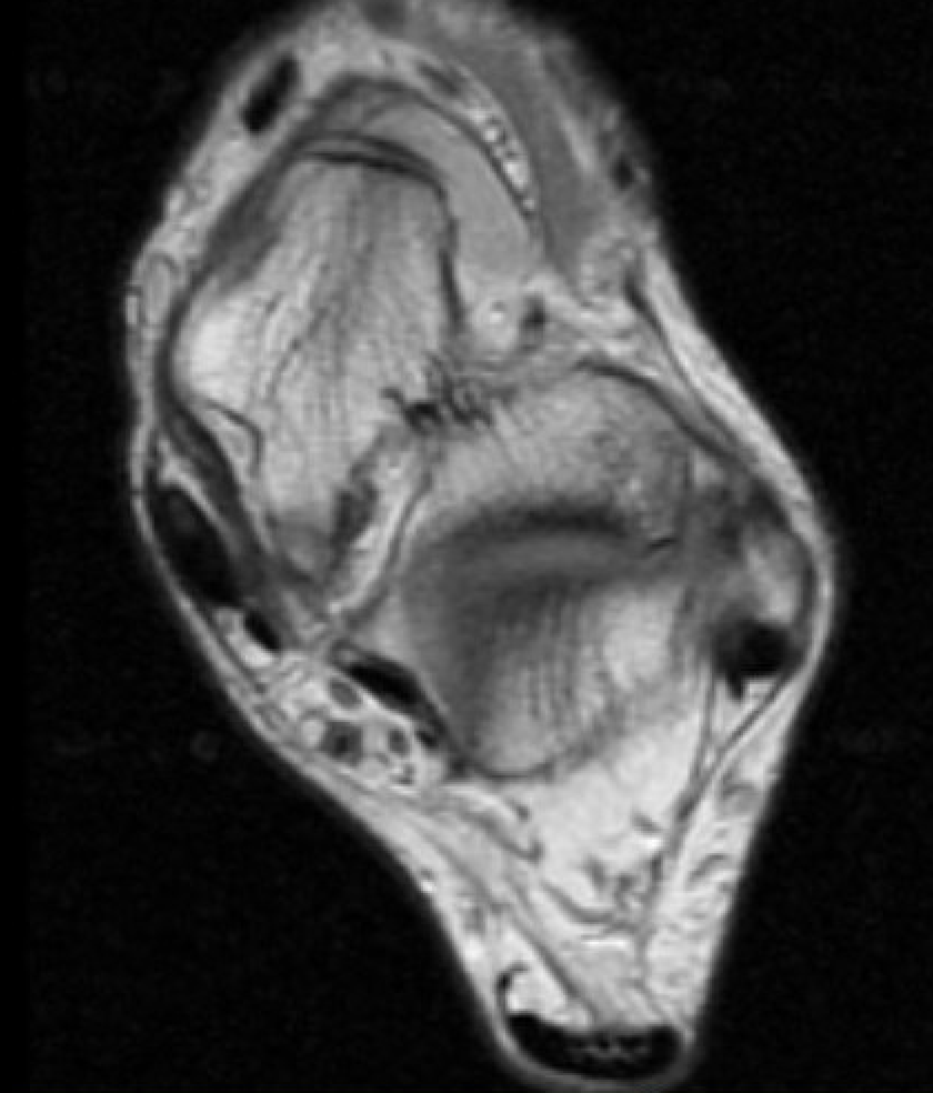
Tibialis posterior tendinopathy
Tear tibialis posterior with 10 cm gap