Johnson Classification
| Stage 1 | Stage 2 | Stage 3 | Stage 4 |
|---|---|---|---|
| T posterior tendonitis | T posterior elongation / attenuation / rupture |
Fixed deformity subtalar joint |
Varus angulation talus in ankle joint |
| Able to single heel raise |
Unable to single heel raise Correctable subtalar joint No subtalar OA |
Non correctable valgus Rigid flatfoot |
|
|
IIA: As above IIB: Forefoot abduction |
+/- subtalar OA | +/- ankle joint OA |
Management options
| Stage 1 | Stage 2 | Stage 3 | Stage 4 |
|---|---|---|---|
|
Orthotics
Tibialis posterior debridement
+/- tendon transfer |
Tendon transfer - FHL / FDL
Spring ligament repair / reconstruction
Calcaneal osteotomy - medial displacement (stage IIA) - lateral column lengthening (stage IIB)
Arthroereisis
Medial column / forefoot surgery |
Double arthrodesis +/- deltoid ligament reconstruction
Triple arthrodesis +/- deltoid ligament reconstruction |
Triple arthrodesis + deltoid ligament reconstruction
Hindfoot or pantalar fusion |
Stage 1 Tibialis posterior dysfunction
Non operative
Walking boot / cast for 4 - 6 weeks
UCBL
- worn inside the shoe
- ends under malleoli
- controls the heel (which must be flexible) & supports the arch
Tibialis Posterior Synovectomy
Technique
Incision
- tip of medial malleolus to navicular
- open tendon sheath
- perform synovectomy +/- repair or tubularization
+/- tendon transfer
Arthroscopy endoscopic technique tibialis posterior debridement PDF
Stage 2 Tibialis posterior dysfunction
FDL tendon transfer
Technique
Vumedi FDL transfer surgical technique video
Incisions
- along entire length T posterior
- 10 cm proximal to medial malleolus
- to metatarsal cuneiform joint
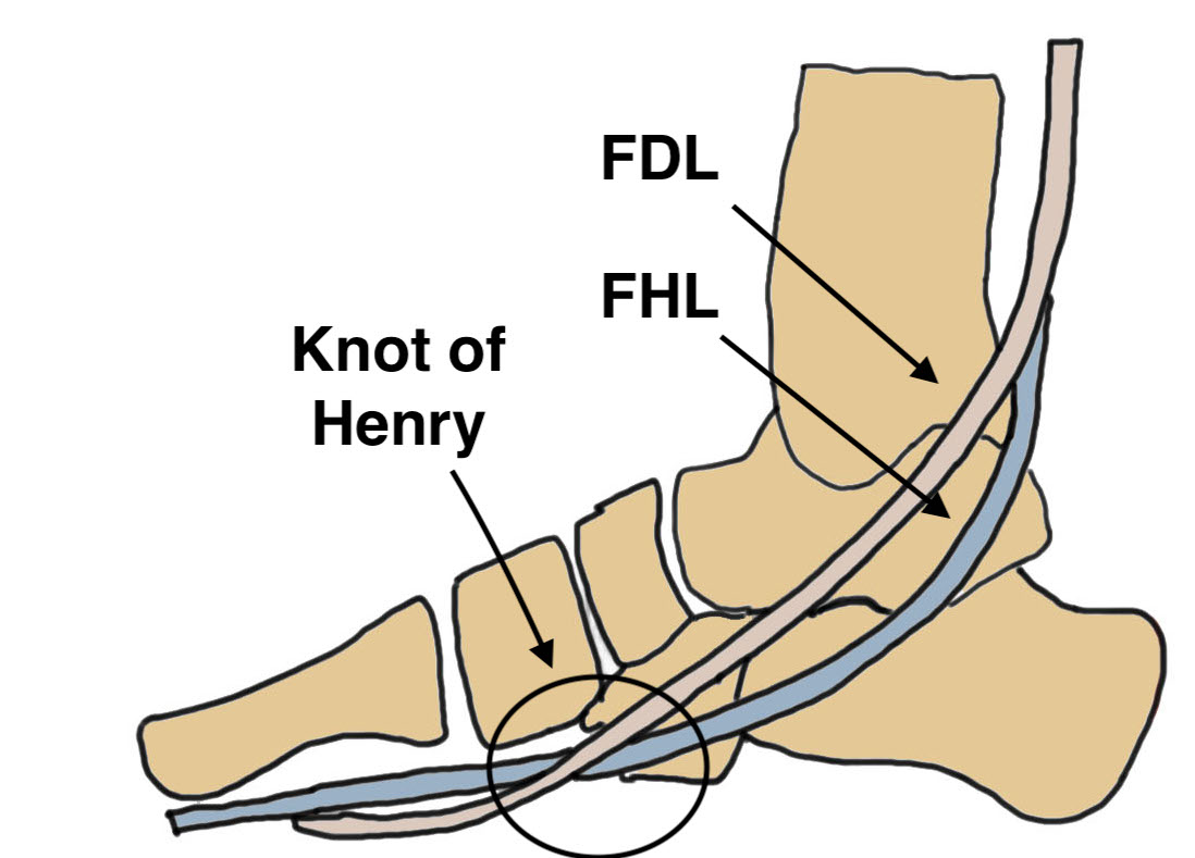
Deep dissection
- T posterior tendon dorsal, divide and reflect abductor hallucis plantar
- Knot of Henry - crossover of FDL & FHL
- FDL plantar to FHL
- suture together and release proximal FDL
Expose navicular
- reinsert FDL into underside of navicular
- plantar to dorsal
- ankle in equinus & forefoot in varus
+/- repair spring ligament / calcaneonavicular ligament
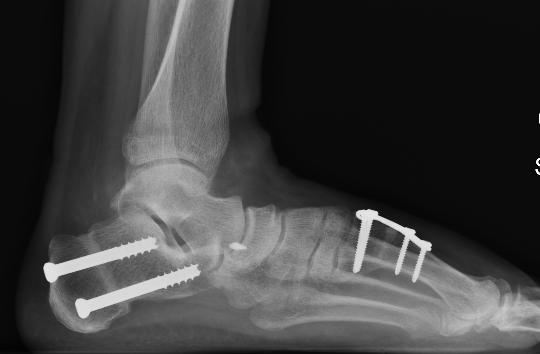
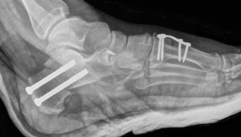
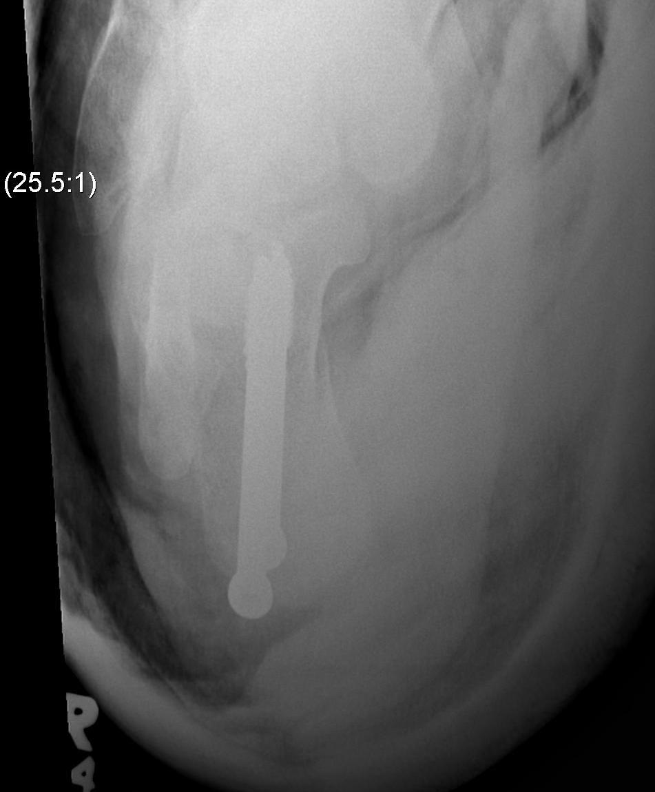
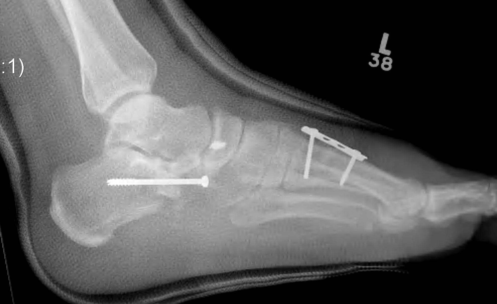
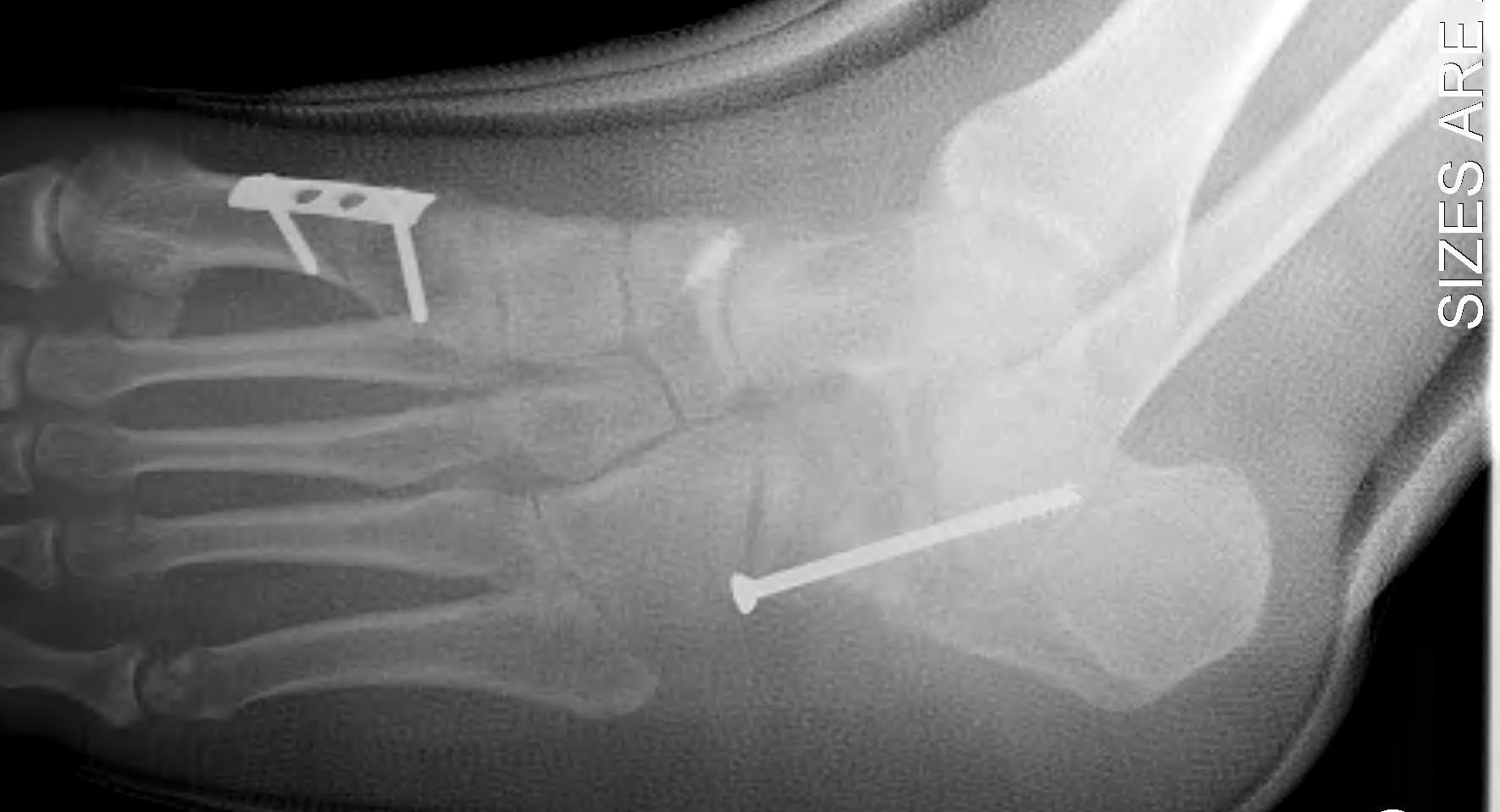
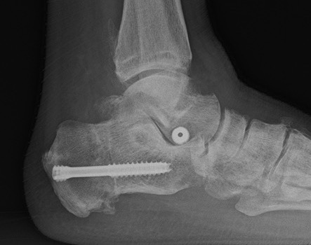
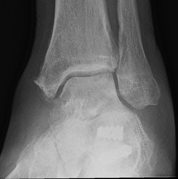
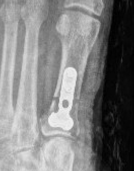
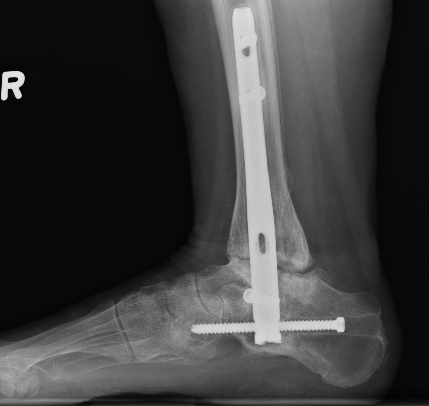
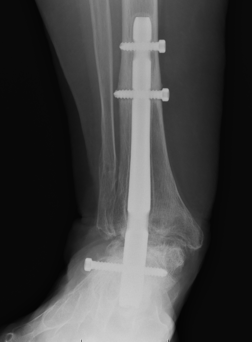
Medial displacement calcaneal osteotomy
Technique
Acumed calcaneal medial shift surgical technique video
Lateral approach
- curve just below peroneals
- protect sural nerve branches
- homann superiorly in front of tendoachilles
- homann inferiorly under calcaneum
Oblique osteotomy behind posterior facet
- 45o cut with saw
- open with lamina spreader
- split periosteum medially with osteotome
- avoid damage to medial structures
- transfer medially 1 cm
- screw fixation
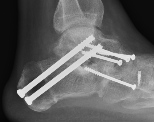
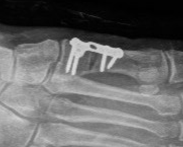
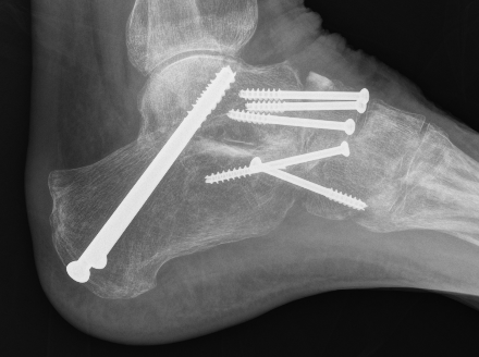
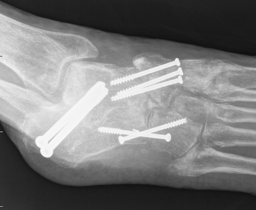
Evans Calcaneal Lengthening Osteotomy
Technique
Vumedi calcaneal lateral lengthening osteotomy video
Incision over anterolateral distal calcaneum
- sural nerve retracted plantar
- P longus retracted plantar
- identify CCJ
- Z lengthen P brevis
- homan retractor in sinus tarsi (between middle and anterior facets)
- homan retractor inferior calcaneum
- K wire into CCJ to prevent subluxation
Opening wedge osteotomy
- 1.5cm proximal to CCJ
- between middle and anterior facets medially
- begin with saw, complete with osteotome
- open 1 cm
- triangular / trapezoidal bone graft (allograft, iliac crest / mid fibular autograft)
- fixation with plate / staple / screw
+/- tendoachilles lengthening
+/- modified Kidner procedure (imbricate spring ligament, Tibialis posterior advancement)
Arthroereisis
Results
Ceccarini et al Foot Ankle Surg 2018
- arthroeresis for 29 stage IIa tibiallis posterior dysfunction
- good / excellent 80%
Silva et al Foot Ankle Surg 2021
- compared lateral column osteotomy v arthroeresis
- 76 patients
- better outcomes with osteotomy and lower complications
- 21% arthroeresis with sinus tarsi pain requiring implant removal
Medial column procedures
Indications
Allow weight bearing of medial column
Forefoot varus
Options
First metatarsal osteotomy
Cotton - opening wedge medial cuneiform ostetomy
First TMT fusion
Stage 3 Tibialis posterior dysfunction / Rigid flatfoot
Options
Double (subtalar / TNJ) versus triple arthrodesis (subtalar / TNJ / CCJ)
- 23 feet with stage 3 T posterior dysfunction
- treated with double v triple arthrodesis
- no difference in outcomes
- shorter operative time with double arthrodesis
Burrus et al J Foot Ankle Surg 2016
- 16 rigid pes planus comparing double and triple arthrodesis
- increased nonunion and worse functional outcomes with double arthrodesis
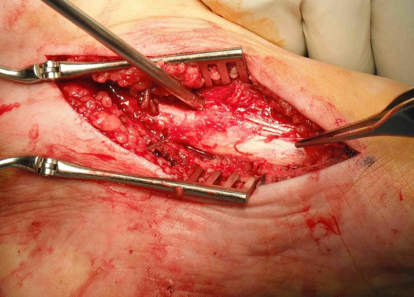
Triple Arthrodesis
www.boneschool.com/triple-arthrodesis
Aim
Realign hindfoot with plantigrade foot
Technique
Vumedi triple arthrodesis cases
Acumedi triple arthrodesis surgical technique video
Lengthen tendoachilles - gastrocnemius recession / release
Lateral approach to subtalar and CCJ / medial approach to TNJ
Remove articular cartilage and prepare joints for fusion
Reduce joints
- Grice maneuver - laminar spreader between anterior process of calcaneus and talus
- check subtalar joint alignment - slight valgus / not neutral or varus
- may need to add medial slide calcaneal osteotomy
Fuse TNJ first to align STJ
Fuse STJ
- may need large lateral bone wedge
- may have issues with lateral skin closure
Fixation - screws / plates / staples
+/- medial osteotomy to allow medial metatarsal weight bearing
- Cotton osteotomy (cuneiform) / first metatarsal osteotomy
Stage 4 Tibialis posterior dysfunction
Hindfoot fusion
www.boneschool.com/pantalar-fusion