Quadriceps muscle tears
British Athletics Muscle Injury Classification (MRI)
Middle age men
Steroids / Growth Hormone
Usually occurs in gym
Bench Press
Significant bruising in the acute phase
In chronic setting, ask patient to adduct against hip / resistance
1. Open antero-lateral approach
Large / Massive Cuff Tear
2. Deltopectoral approach
Large Subscapularis tear
3. Arthroscopic Assisted Mini-open
Indication
- Small / Moderate Cuff Tear < 3cm
- no retraction
Technique
- arthroscopic SAD
Pain & Stiffness
- often more pain than FT tears
Bursal side tears more painful than articular
Articular side more common
May see in young patient overhead throwing
Painful arc
Impingement signs
No weakness
- function good
Largest and most powerful rotator cuff
- arises coastal border of scapula
- superior 2/3 tendon inserts into LT
- inferior 1/3 inserts into proximal humerus
Action
- IR (with T major, P major, Lat Dorsi)
- part of force couplet depressing humeral head
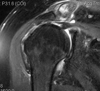
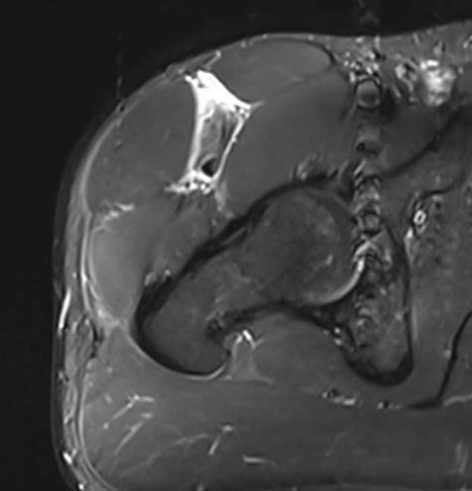
Massive tear
1. > 5cm
- retracted to humerus / glenoid margin
2. At least 2 complete tendons
- lose SS / IS or SS / SC
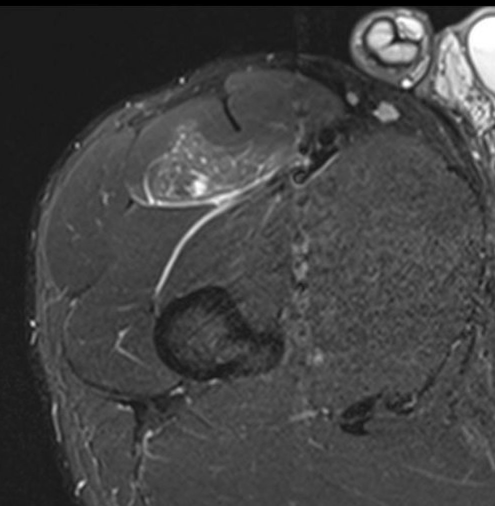
Full thickness tear (FTT)
- variable amount retraction from insertion
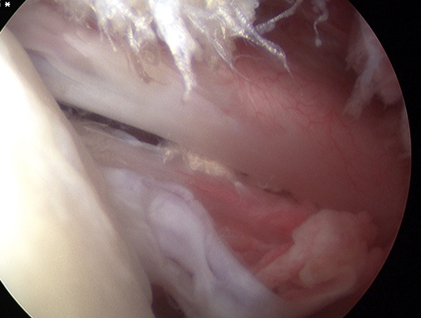
Partial thickness tear (PTT)
- incomplete
- bursal or articular sided
Lateral compartment of leg
- run through retromalleolar groove
- pass superior and inferior to peroneal tubercle
- covered by inferior peroneal retinaculum
Peroneus longus
- origin lateral condyle of tibia and head fibula
- tendon PL superficial and inferior to brevis in retromalleolar groove
- runs in cuboid groove
- insert plantar surface base of 1st MT and lateral aspect medial cuneiform