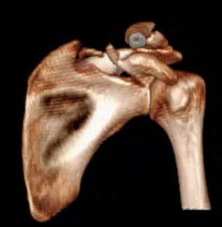
Definition
Intra-articular scapular fracture
Epidemiology
Scapular fractures 1% of all fractures
Glenoid fractures 20% of scapular fractures
Younger patients with high velocity injuries - MVA, fall from height
Older patients with anterior shoulder dislocations
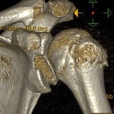
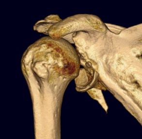
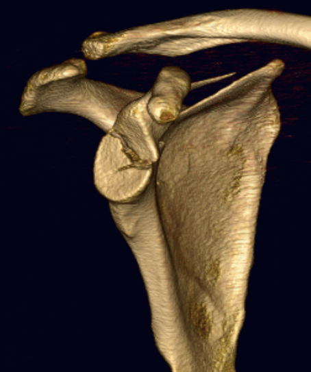
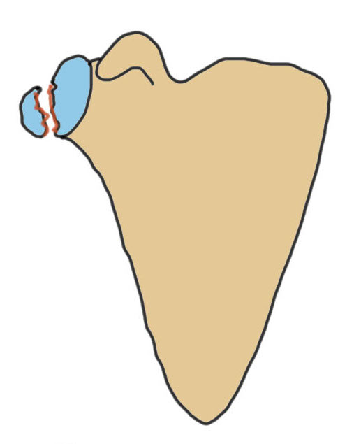
Ideberg Classification Intra-Articular Fracture
| Type IA | Type 1B | Type II | Type III | Type IV |
|---|---|---|---|---|
|
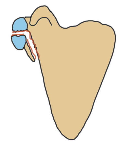
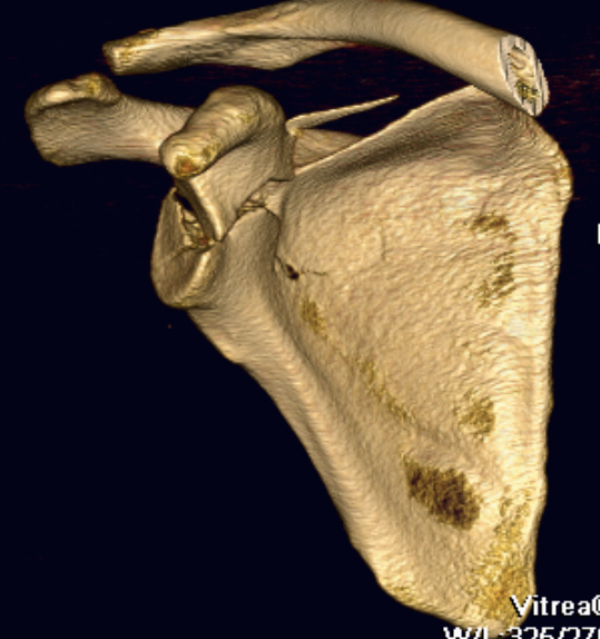
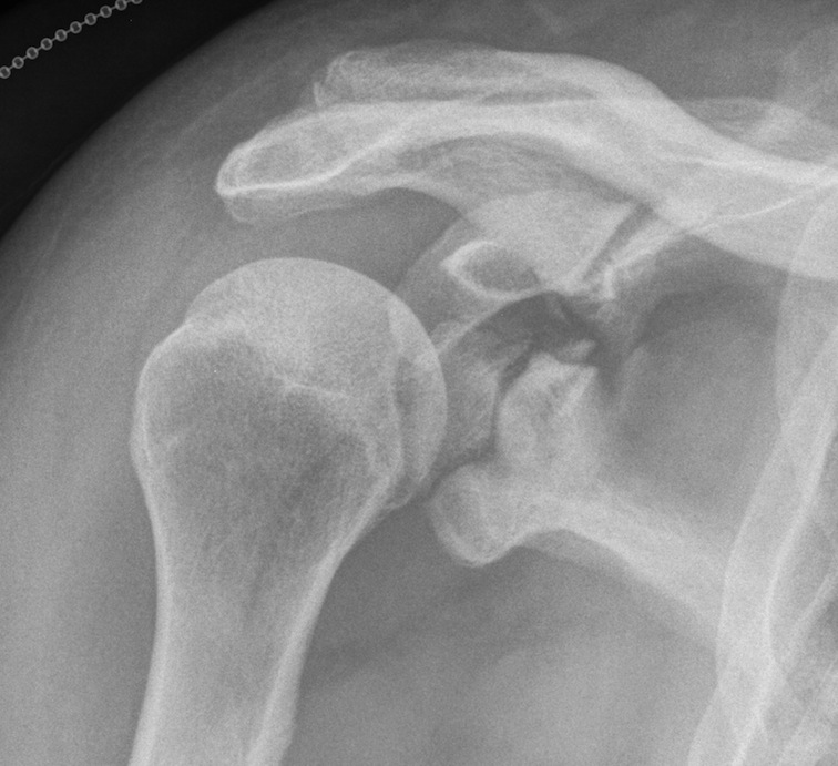
Anterior glenoid rim #
|
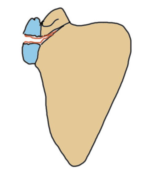
Posterior glenoid rim #
|
Fracture glenoid fossa Exits laterally Inferior fragment |
Fracture glenoid fossa Superior fragment |
Fracture glenoid fossa Exits medially |
|
|
 |
 |
 |
 |
Type V: combination of Type I, II, III & IV
Type IV: severe comminution
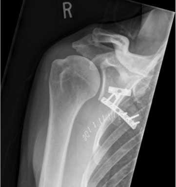
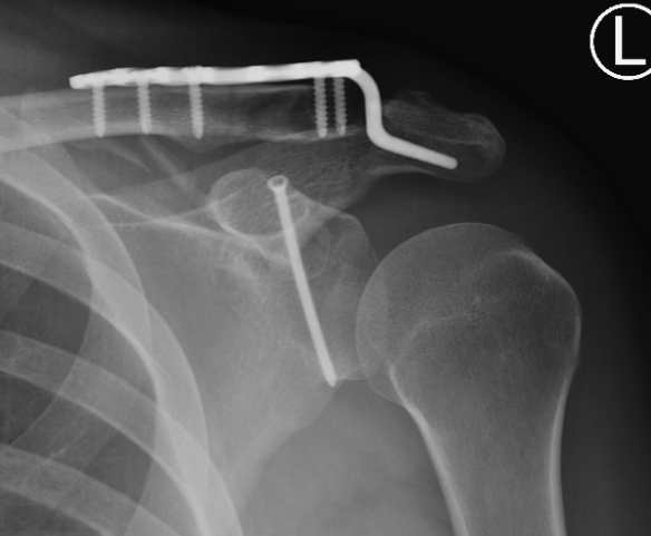
Operative indications
> 20 - 25% glenoid fossa
Displacement > 4 - 5 mm
Humeral head subluxation
Associated scapular fractures
Floating shoulder
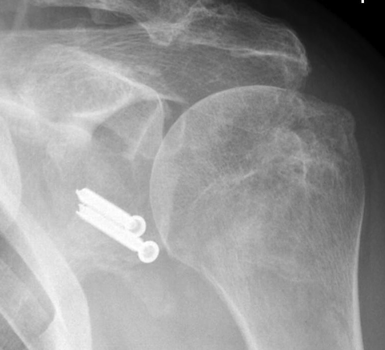
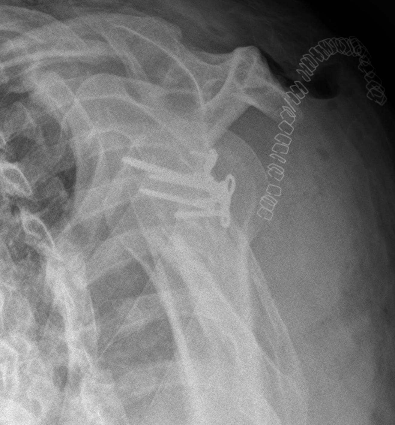
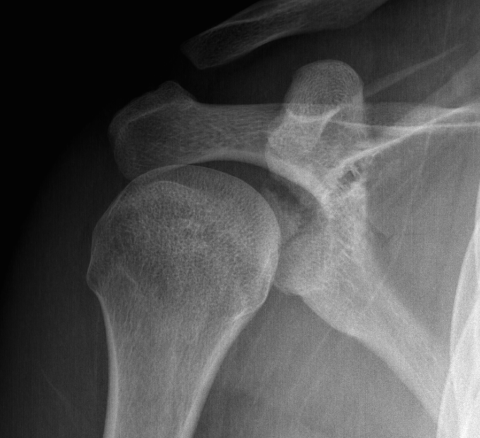
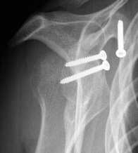
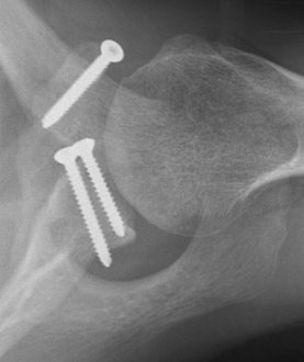
Type IA: Anterior glenoid rim fracture
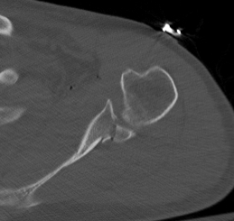
Xray / CT
Technique
Options
Open / Arthroscopic
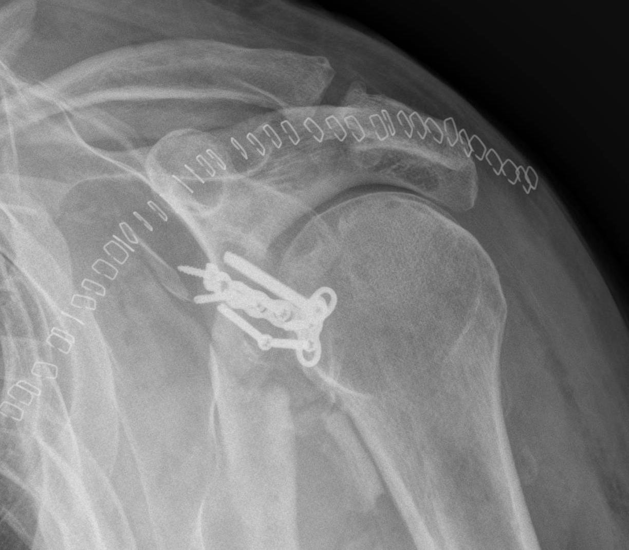
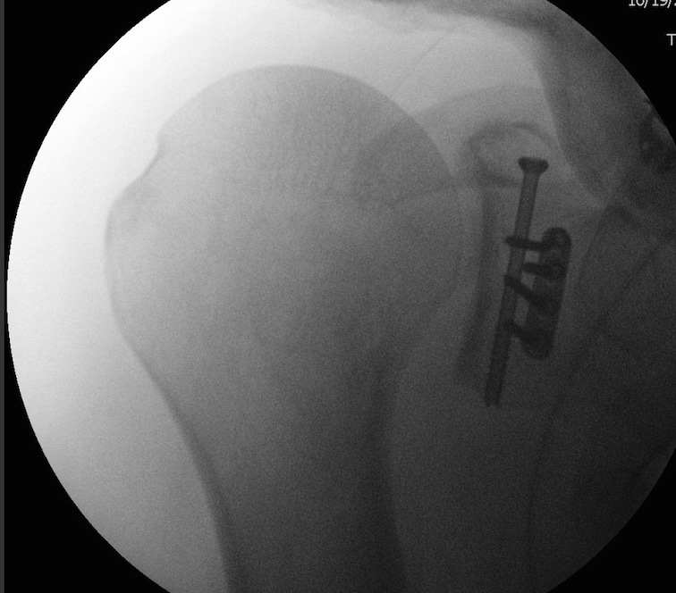
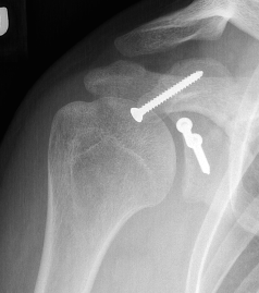
Screw / anchor fixation - depends on size / comminution
Open
Deltopectoral approach
- subscapularis split / subscapularis takedown
- capsulotomy - view articular reduction
- screw / Anchor fixaiton
Arthroscopic
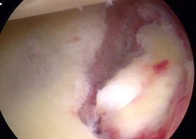
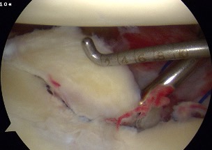
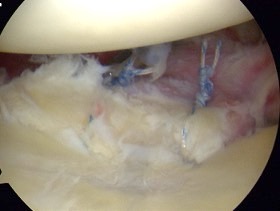
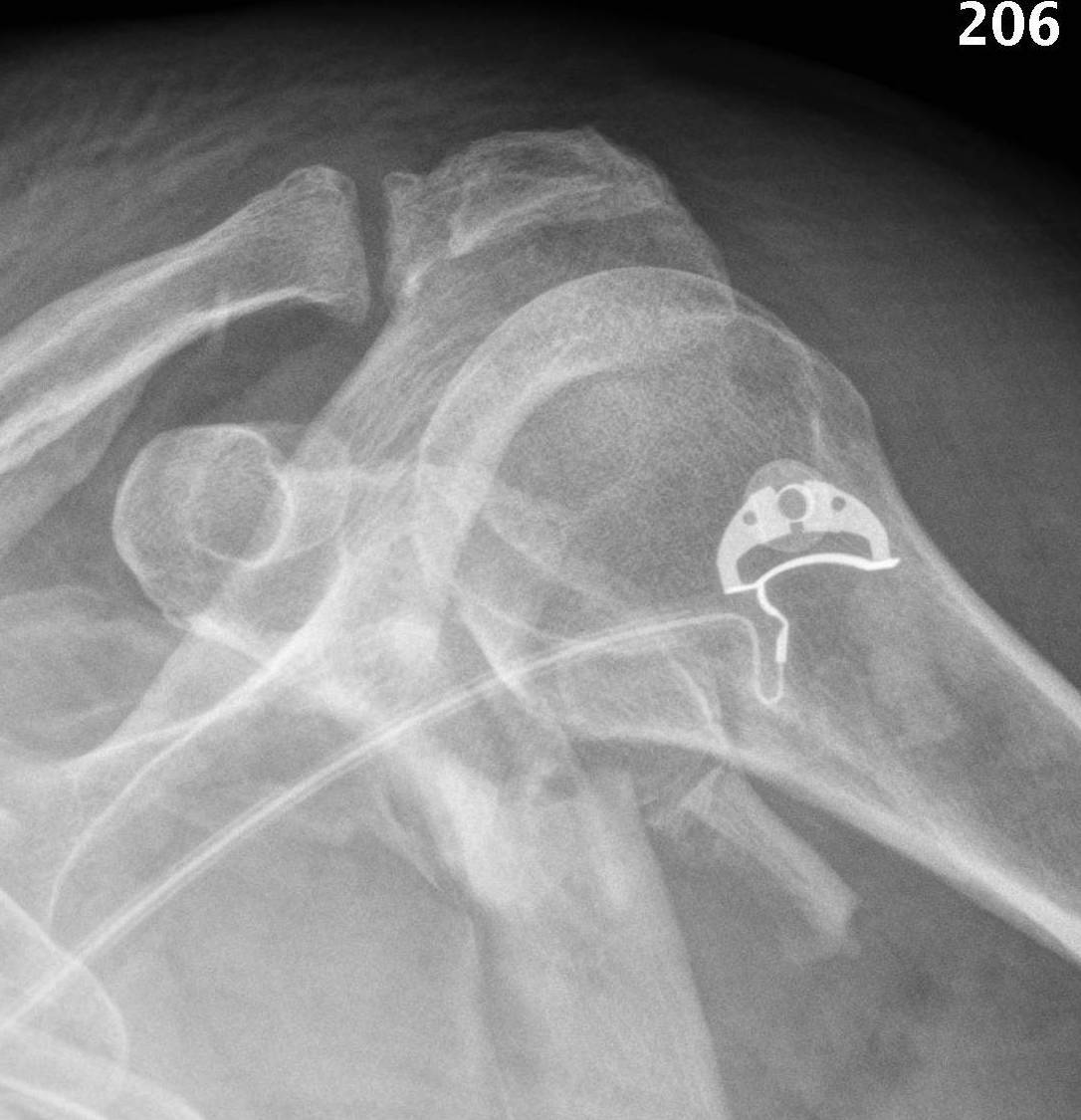
Arthroscopic suture anchor fixation anterior glenoid fracture
Anchor fixation
Vumedi arthroscopic anchor fixation glenoid fracture video
Arthroscopy techniques anterior glenoid rim arthroscopic anchor fixation PDF
Screw fixation
Vumedi arthroscopic screw fixation glenoid fracture video
Arthroscopy techniques anterior glenoid rim arthroscopic screw fixation PDF
Results
- 50 anterior bony bankart arthroscopic repairs with suture anchors
- 95% return to sport
- 7% redislocation
- 17% nonunion - more common with chronic cases
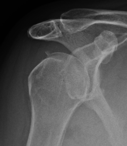
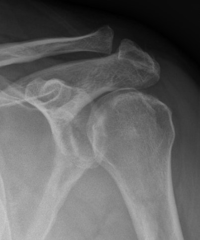
Type IB: Fractures of the posterior glenoid rim
Technique
AO surgical foundation posterior approach glenoid / scapula
Brodsky / Posterior approach to glenoid
- incision from posterolateral acromion
- elevate deltoid
- Window 1: interval between infraspinatus and teres minor
- Window 2: interval between supraspinatus and infraspinatus
- Window 3: interval beween teres minor and major / circumflex scapular
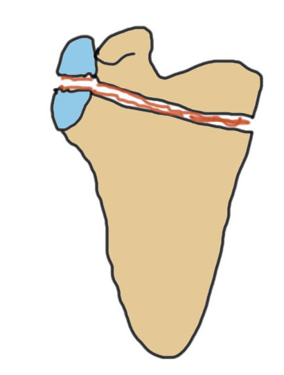
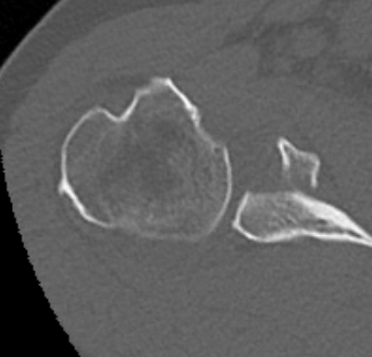
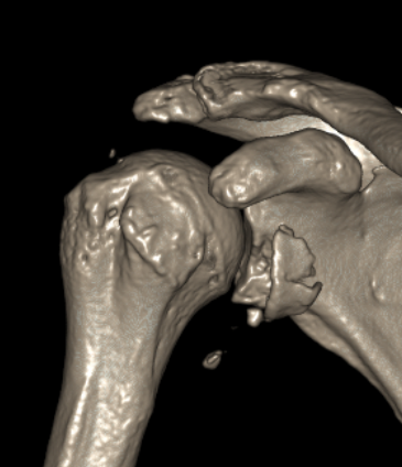
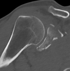
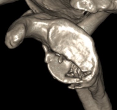
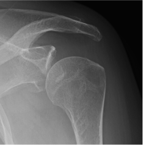
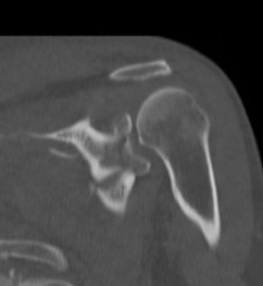
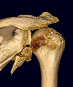
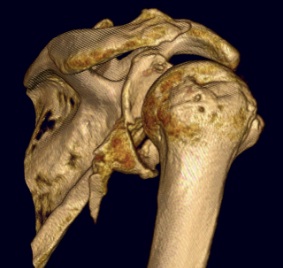
Type II: Fracture glenoid fossa that exits laterally
Definition
Transverse fracture through glenoid fossa
- inferior triangular fragment
- exits lateral border scapula
![]()
Technique
Options
Posterior approach - Judet / Brodsky / posterior based fractures
Direct / trans-axillary approach
AO surgical foundation posterior approach glenoid / scapula
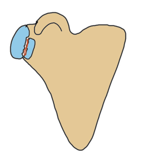
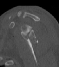
Type III: Fracture of the glenoid fossa that exits superiorly
Definition
Fracture through the glenoid fossa exiting through superior border of the scapula
- superior fragment
- includes the coracoid
Technique
Options
Open - anterior approach +/- coracoid osteotomy, superior approach (suprascapular fossa)
Arthroscopic
Vumedi arthroscopic fixation Type III fracture video
Arthroscopy techniques arthroscopic fixation Type III fracture PDF
Results
- 23 Ideberg Type III treated with arthroscopic screw fixation
- no hardware failure or redisplacement
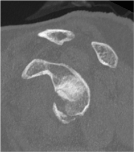
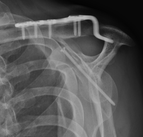
Type IV: Fracture of the glenoid fossa exiting medially
Definition
Similar to Ideberg Type III
- larger superior fragment
- horizontal and exiting thru the medial border of the blade